
This post was last updated on September 16, 2026.

The following is a long summary of my research notes and some self-experimentation that I engaged in as I waited for an appointment with a mast cell specialist, as well as updates from that appointment. It is an aggregate of my own digging and learning, shared for information and education purposes and not a substitute for specialist care.
A SUMMARY OF IMPORTANT POINTS
The following are discussed in more detail on this (long!) page. A summary of some main points:
– Mast cells exist all over the body, and symptoms can therefore occur all over the body. I am celiac, but my MCAS does not manifest with many GI symptoms, perhaps because I’ve long had that aspect under control. Other patients I know are constantly in the bathroom. It will differ person-to-person, and one patient may have anaphylaxis that differs from another patient’s symptoms and reactions.
– For patients with abnormal connective tissue due to Ehlers-Danlos Syndrome or another connective tissue disorder, significant periods of mast cell dysfunction may result in visible changes to skin. With abnormal mast cells and collagen synthesis, and with mast cells living in the body’s tissues, this seems to be the most common comorbidity in the patient cohort. In noting “significant periods”, I include clearing viruses like EBV, SARS-CoV-2 and dengue fever, among others.
– With hundreds of mediators released by mast cells, affecting many receptors in the body, there’s much to look at. However, tests have yet to be developed to measure many of them. Each patient’s symptoms will be different, usually with some overlap. Histamine is often discussed in literature or blog posts online, but that is insufficient. There are far, far more mediators released than just histamine, and it’s important to not ignore the others.
– This also means that triggers can be different for each patient, be it environmental triggers or food-related triggers. These include excipient ingredients (fillers) in pills, radiological dyes and even procedures that cause pain. It is important to premedicate for procedures and surgeries. More recently, scientists have also identified that the MRGPRX2 receptor, located on mast cells and on certain neuronal cells, may be responsible for hypersensitivity to different medications, as well as reactions to triggers like contrast media in imaging like CT or MRIs. Research is fairly new in this area, and for now avoidance of triggers remains the bottom line. See the recommendations under “triggers”, below. Some people even react to their own sweat (I know, right?), or to vibrations, or to sunlight. One common trigger for most patients is situations of trauma and stress. The many other triggers that exist will vary person to person.
– A mast cell flare is not the same as a “true” allergy. True IgE allergies can be identified with blood or skin tests. Mast cell reactions vary based on someone’s baseline, and are not identifiable in the same way. See the bucket analogy graphic, below. A patient can have “true” allergies alongside MCAS, however.
– Many conditions are comorbid (commonly occur with) with mast cell disorders, some of which are set out on this page. Others can be mediated by mast cells, even if not a documented comorbidity.
–Diagnosing mast cell conditions is not simple. Many practitioners erroneously believe that a normal tryptase is conclusive that the patients have no mast cell disorder. There is some dispute among mast cell experts about what measures are necessary to diagnose the condition; the details are set out below. Also, patients sometimes get diagnoses (MS, lupus, anxiety, interstitial cystitis and more) that reflect one arm of mast cell problems, only to find out later that treating MCAS goes a long way to mitigating those (and other) symptoms. You may want to print this 2-page PDF about MCAS created in 2026 for you to share with your physicians, courtesy of nonprofit Mast Cell Action.
– Given how patients differ with respect to triggers, it makes sense that treatment is also bioindividual. Baseline treatment for mast cell activation syndrome is usually an H1 and an H2 antihistamine taken together, twice a day. This 12-hours-apart regimen is known by some experts as a “histamine blockade”. Finding the types of medication and brands that work best for you will involve testing different options methodically until landing on one your body likes. It’s tedious, but necessary. In addition, many patients benefit from natural mast cell stabilizers like quercetin, as set out below in the “treating naturally” section. Some patients also require prescription mast cell stabilizing medication, like ketotifen or sodium cromolyn. I have shared what worked for me thus far, but given how complex and varied the condition is, it does not favour a one-size fits all approach.
– Treating mast cell activation syndrome with only natural products is a difficult ask for some patients. I find it problematic that some blogs or sites shame patients for using a combination of antihistamines and natural products. The reality is that while working with underlying causes is important, for many patients doing so will STILL require a stabilization beyond what natural products can do. I have heard from patients only using natural products who then have to add H1/H2 antihistamines, and feel shame about it. This is unacceptable. I have not met a complex patient who was “lazy” about their health; everyone wants to get better and is willing to work hard to get there. Using antihistamines does not mean a patient has “failed”, it means that their root cause is hard to come by (genetics, for some!), and science has yet to catch up with their body’s needs. I do think lifestyle changes including diet, neural retraining, EMDR, supplements, and more are critical. But uncontrolled inflammation is no good for anyone, and if you are a patient where natural options alone do not suffice, do not let anyone shame you for it. (For what it’s worth, many naturopathic and functional doctors also discuss the need for pharmaceuticals over and above natural stabilizers and treatment.)
– Speaking of the science: as of right now, there is currently no established curative treatment for MCAS; management focuses on controlling symptoms, identifying relevant triggers or underlying conditions, and treating associated disorders. There are some new receptor targets that look promising. And there are some cases where root causes are found, e.g. some practitioners have found underlying immune deficiency where treating that helps mitigate the issue. Patients sometimes take this to mean the person is back to ‘normal’, so to speak. While I do hope the science evolves to find a way to put the genie back in the bottle, even in those cases anecdotally a baseline treatment is still required a lot of the time. But be wary of people (especially paid-courses!) that boast “curing MCAS”. For those without a full-blown mast cell disorder, lifestyle changes like diet and supplements and more may resolve their issues. With MCAS and sister disorders, however, there is only management and temporary remission.
– Some mast cell experts are looking at the condition as an epigenetic, and not only a genetic disease, meaning that it would in principle be reversible with the right medication targeting the epigenome. There’s a paucity of research in this area for now, but it is an interesting avenue to keep an eye on, what an April 2022 paper called the “novel concept” of mast cell diseases being rooted in an unnamed transgenerationally transmittable epigenetic disease.
– Many patients with anxiety have found that treating mast cell activation goes a long way toward lowering their baseline anxiety levels. This is not everyone, but specialists have documented cases where patients who previously had panic attacks, prolonged insomnia, and even a diagnosis of generalized anxiety disorder have watched those disappear with treatment for MCAS. In my case, I always had trouble sleeping and thought I had anxiety. Both of these problems went away now that I have my mast cells under more control.
– With Long Covid / PASC in the mix during the pandemic, some mast cell specialists are seeing an explosion of MCAS in that group of patients. I do hope the seeming correlation between Long Covid and mast cell-mediated hyperinflammation leads to more research that would benefit us all.
– And finally (long summary for a long page, I know), a newer condition called Hereditary Alpha Tryptasemia (HαT) is something patients with higher tryptase ought to look into. While MCAS testing is more complicated, HαT testing is a simple DNA test that any doctor—PCP/GP or specialist—can request via a company called Gene by Gene (see link below under diagnosis). HαT occurs where patients have inherited extra copies of the alpha tryptase gene (TPSAB1), and this leads to increased levels of tryptase protein detected in the blood, whether a reaction is happening or not. Per the research, the more copies one inherits, the higher the blood tryptase level. Unlike MCAS, this is easily documented and useful to rule in or out. While treatment is not too different, it will likely result in physicians taking the condition more seriously since testing is so straightforward.
MY MAST CELL STORY IN BRIEF
For me, the first sign of these issues was actually in childhood. I had a cough that would itch my throat when trying to sleep, causing sleep disturbance from a young age. The cough went away when I hit puberty, but came back when I got sick on the trans-Mongolian trains en route to Ulaanbaatar. Then, following dengue fever in Vietnam, more hints: itchy skin when I exercised, itching when I ate certain foods like blue cheese, or drank red wine, and a first foray into seasonal allergies during my first spring in Oaxaca, Mexico. They were so bad that my eyelashes fell out.
I had no idea what was going on, and only ended up looking at mast cells as a potential cause after I sustained a spinal CSF leak following a lumbar puncture in 2017. I saw many leak patients dealing with very similar issues, read up on dengue and mast cells (who knew!) and then it all culminated in my going into anaphylaxis during my 4th blood and fibrin glue patch and needing an epinephrine jab on the table. I have been working on managing my ‘angry’ mast cells since that fibrin glue patch in early 2018.
For reference, prior to starting the regimen below, I had 24/7 searing/burning pain on my skin (spine, hands, feet, etc), adrenaline dumps at night that kept me up until 5am with my heart racing, I’d shake from cold exposure or cold drinks, low blood pressure, hives, gastrointestinal distress, and anaphylaxis on exposure to certain triggers. Since using the list of supplements/meds that I shared here, those have all disappeared, save for occasional reactions to new foods or smells. The skin burning disappearing was especially a relief, as was my ability to sleep again.
I update this page often, and welcome corrections or additional research. Please use the contact page on the menu bar above to send me an email.
This is a free page, but support from my readers helps me continue to create resources pages like this one. I’ve shared a one-time support option below, or you can join my Patreon, where I also provide resources for living with chronic pain.

STARTING AT THE BEGINNING: WHAT ARE MAST CELLS?
Mast cells existed before mammals’ adaptive immune system developed, some 500 million years ago, but modern research only began looking into them in the late 1800s. In 1878, Paul Ehrlich was a 24-year-old medical student in Leipzig where he described a cell full of tiny ‘granules’ and named them Mastzellen, or mast cells. Granulated connective tissue cells had been seen before him by Friedrich von Recklinghausen in 1863, but Ehrlich was the one who stained them and gave them a name and a description. In German, one definition of the word ‘mast‘ is that it relates to the fattening of animals; because these cells appeared to be full of little particles he called them ‘fattening cells’, thinking that they pulled nourishment from the areas around them.
Only in the mid 1900s did medicine discover that mast cells released histamine, one of the many mediators described below — but the most well-known one. In excess, histamine can lead to a spectrum of reactions, from reactive sneezing or eye watering to anaphylaxis. Our body is full of mast cells, and they have enough histamine to cause serious havoc for us. But it took scientists until fairly recently to begin unravelling the mystery of what those cells do and how they can be managed when they are dysfunctional. There is still much to learn.
Mast cells are the sentinels of our immune system, derived from bone marrow progenitors (CD34+ cells). They sit on our immunological front line to help control the immune processes in the body. They are called resident immune cells because they reside in tissues and not in our bloodstream. And they reside in tissues all over the body, from the connective tissue, to the endothelial cells, to the epithelial cells, and even in the brain.
Mast cells are immune system cells that live in the bone marrow and in body tissues, internal and external, such as the gastrointestinal tract, the lining of the airway, and the skin. Everyone has mast cells in their body, and they play many complex and critical roles in keeping us healthy. The positive roles that they play include protecting us from infection, and helping our body by participating in the inflammatory process. However, mast cells are also involved in allergic reactions, from the tiny swelling that appears after a mosquito bite to a life threatening, full-blown anaphylaxis.
-The Mast Cell Disease Society, formerly The Mastocytosis Society of the United States (TMS); Source.
Mast cells are important to the immune system; we need them! When mast cells are acting ‘normally’ in our bodies, the reactions we have to allergens or triggers are our bodies’ attempts to remove these triggers from our body and then instruct our immune system to avoid them going forward. But, when they are behaving dysfunctionally, as is likely the case if you’re reading this article, mast cells can react to the most innocuous of triggers, requiring significant stabilization to improve quality of life.
Dysfunction of mast cells may lead to severe diseases, and can lead to a wide variety of problems and symptoms. Since the mast cells reside all over the body, those symptoms are often systemic. And since each of us has a different immune system, the symptoms and manifestations of mast cell disease vary across the patient population.
Mast cell degranulation and other mast cell secretion of mediators
As noted above, mast cells are filled with tiny granules, as you can see in the screenshot of the video below. When they get “angry,” a destabilization process known as degranulation, they dump hundreds of different signalling chemicals — called mediators — into the bloodstream. Those mediators are inflammatory, and the degranulation process causes a cascade of inflammation that can affect other workings in the body. Mast cells exist in organs and tissue throughout the body, and mediate allergic, immune, and inflammatory reactions.
We don’t want to eliminate the mast cell’s ability to degranulate. As the quote above illustrates, the degranulation response is part of the body’s natural defence when it senses invaders, and part of what helps keep us safe and healthy.
The problem is that in mast cell activation syndromes, that normal defence response goes off the rails. The mast cells get riled up* and mount a defence for innocuous things that would not otherwise be a threat.

Essentially, the normal mast cell process is altered and the mast cells are sensitized and degranulate with exposure to a variety of triggers, including excipients in medication, stress, and more. I go into the types of triggers below.
*Yes, I am anthropomorphizing mast cells. I find it helpful to think of them as friends I’m trying to negotiate with.
Below is a short video showing what happens when mast cells degranulate:
And here’s an image, for those who don’t have the bandwidth for video:

In addition to what mast cells degranulate, recent studies have focused on how the mast cells degranulate. And I don’t mean triggers — that information is below — but rather that there are different means that mast cells dump mediators; it’s not just a wholesale degranulation each time.
A 2024 article by Dr. Theoharis C. Theoharides called Mast cells: The unregulated master immune response conductor discusses how mast cells can secrete mediators in several different ways and pathways, including via
- degranulation (covered above)
- compound exocytosis, where multiple secretory granules fuse with each other and then with the plasma membrane of a cell, releasing their contents in a massive, coordinated manner.
- piecemeal degranulation.
- differential release
- extracellular microvesicles, small membrane-bound vesicles released by cells (including mast cells) into the extracellular space, acting as messengers in cell-to-cell communication; and
- nanotubules (proposed intercellular structures)
He used the following image to graphically illustrate these secretion means:

WHY DO MAST CELLS GET DYSFUNCTIONAL?
Types of mast cell diseases
This is the million dollar question, and one scientists are still examining today. Depending on the type of mast cell disorder, the mast cells may proliferate from bone marrow in excess (mastocytosis), or react dysfunctionally to previously tolerated triggers (mast cell activation syndrome) or have a genetic basis for behaving unusually.
We do know that mast cell diseases are caused by the proliferation and accumulation of sensitized or altered mast cells, or the inappropriate release of mast cell mediators, creating symptoms in multiple organ systems. There are several main forms of mast cell diseases: mast cell activation syndrome, mastocytosis, and the newer Hereditary Alpha Tryptasemia Syndrome (HαT). Mastocytosis is further broken down into different subtypes. There is also mast cell leukemia, a rare and aggressive subtype of systemic mastocytosis.
Mast cell diseases can cause tremendous suffering and disability due to symptomatology from daily mast cell mediator release, and/or symptoms arising from infiltration and accumulation of mast cells in major organ systems. Although systemic mastocytosis is a rare disease, those suffering with MCAS have recently been increasingly recognized and diagnosed. As a result, patients with MCAS appear to represent a growing proportion of the mast cell disease patient population. It is important to note that the process of mast cell activation can occur in anyone, even without a mast cell disease, as well as in patients with both mastocytosis and MCAS.
– TMS
MCAS, HαT, Mastocytosis, and Histamine Intolerance
The focus of this page is mast cell activation syndrome (MCAS), which is when the mast cells are hyperactive and degranulate easily, and too frequently. An April 2020 study estimates that “this disease that could affect up to 17% of the population on a spectrum from very mild to debilitating symptoms. MCAS is often either misdiagnosed or the diagnosis is greatly delayed due to a lack of provider awareness.” While population prevalence is not established, we do know that MCAS is often misdiagnosed, or diagnosed very late, because so few providers are aware of it. Harbingers of MCAS are often present in a patient’s life before the condition fully emerged, such as the clues from childhood mentioned above.
In mastocytosis, a patient has too many mast cells and they are often also prone to easy degranulation. Mastocytosis, of which there are several varieties, is often confirmed with a bone marrow biopsy that looks for a KIT gene mutation. This is because more than 90% of patients with mastocytosis possess mutations in the KIT gene, most of them in KIT D816V.
There are several different variants of mastocytosis, but most common are cutaneous mastocytosis, and systemic mastocytosis. Diagnosis for mastocytosis is often as follows:
- Cutaneous mastocytosis is diagnosed by the presence of typical skin lesions and a positive skin biopsy demonstrating characteristic clusters of mast cells.
- Systemic mastocytosis is usually diagnosed via bone marrow biopsy. The WHO criteria were updated in the 5th edition of its classification in 2022. They combine a major criterion based on dense multifocal infiltrates of mast cells with minor criteria that include the KIT D816V mutation, abnormal mast cell morphology, aberrant surface markers, and elevated serum total tryptase. Note that extra copies of TPSAB1 as found in Hereditary Alpha Tryptasemia can raise baseline tryptase on their own, even in the absence of systemic mastocytosis.
Then there is Histamine Intolerance. My mast cell specialist considers histamine intolerance to represent a milder form of mast-cell-related dysfunction; other clinicians regard it as a distinct and still incompletely defined condition, potentially caused by microbiome or intestinal imbalances, or genetic issues that create a lack of Diamine oxidase (DAO) in the body. DAO is an enzyme that helps break down histamine. The DAO and HNMT pathways are both implicated in histamine intolerance, and certain medications and foods can deplete DAO in the body as well. Per some physician experts in the field, taking supplemental DAO and going on a low histamine diet can help these patients — see my recommendations in the “treating naturally” section below.
That said, mast cell diseases transcend histamine alone; with hundreds of mediators released by the mast cells, histamine is but one of them. For those where DAO and low histamine diets are insufficient, you may want to think about in-depth mast cell testing.
This image below from a January 2020 editorial entitled “Mast Cells: Fascinating but Still Elusive after 140 Years from Their Discovery,” is also a helpful pictorial demonstration of the many conditions that can arise when the normal mast cell process gets dysfunctional.

The same study notes:
During the last years, it became evident that mast cells represent an important cell during bacterial, fungal, viral, and helminth [a type of parasitic worm] infections. Elegant studies have demonstrated that mast cell-derived mediators can play protective roles against several venoms. Mast cells and their mediators can be involved in several aspects of tumor initiation and growth, presumably through the production of several angiogenic and lymphangiogenic factors. Systemic mastocytosis is a clonal disease associated with a somatic gain-of-function KIT mutation. Mast cells, strategically located in different sections of the human heart and atherosclerotic plaque, are involved in different phases of atherosclerosis and myocardial infarction. These cells can be involved in several autoimmune disorders, such as rheumatoid arthritis, coeliac disease, multiple sclerosis, and bullous dermatoses.
EXPLAINING MCAS TO FAMILY AND FRIENDS
One of the challenges in having a systemic, complex disease is how you communicate your needs and/or explain the condition to family and friends.
Most people are familiar with traditional allergies, which are described below and consist of an allergen that is a constant trigger, often worsening each time you are exposed. Given the systemic aspect of mast cell disease, it’s less binary and less “I’m going to die if I come into contact with this thing” — though there are mast cell patients who also have IgE allergies. For example, I am allergic to bee venom and have an EpiPen for it. This is a non-changing allergy that is very severe in its anaphylaxis and I have to be vigilant.
Unlike IgE allergies, living with MCAS feels like doing a twisted form of calculus all day long to avoid ‘filling your bucket’ and leading to prolonged reactions. Can you eat this thing? Well, how stressed are you right now? How much pollen is in the air? How many mast cell stabilizers are you taking?
It’s a lot of spinning plates to keep track of, but once you stop trying to make binary rules and let yourself explore the triggers you may have with curiosity instead of resentment, it becomes a lot easier to keep track of. Some things may always cause a reaction, but others may be only something you react to in tandem with how full your ‘bucket’ is.
I highly recommend getting a notebook to keep track of what you eat, supplements or medication you’re taking, what you’ve changed in your environment, and your symptoms to better allow you to narrow down on what your specific triggers are.
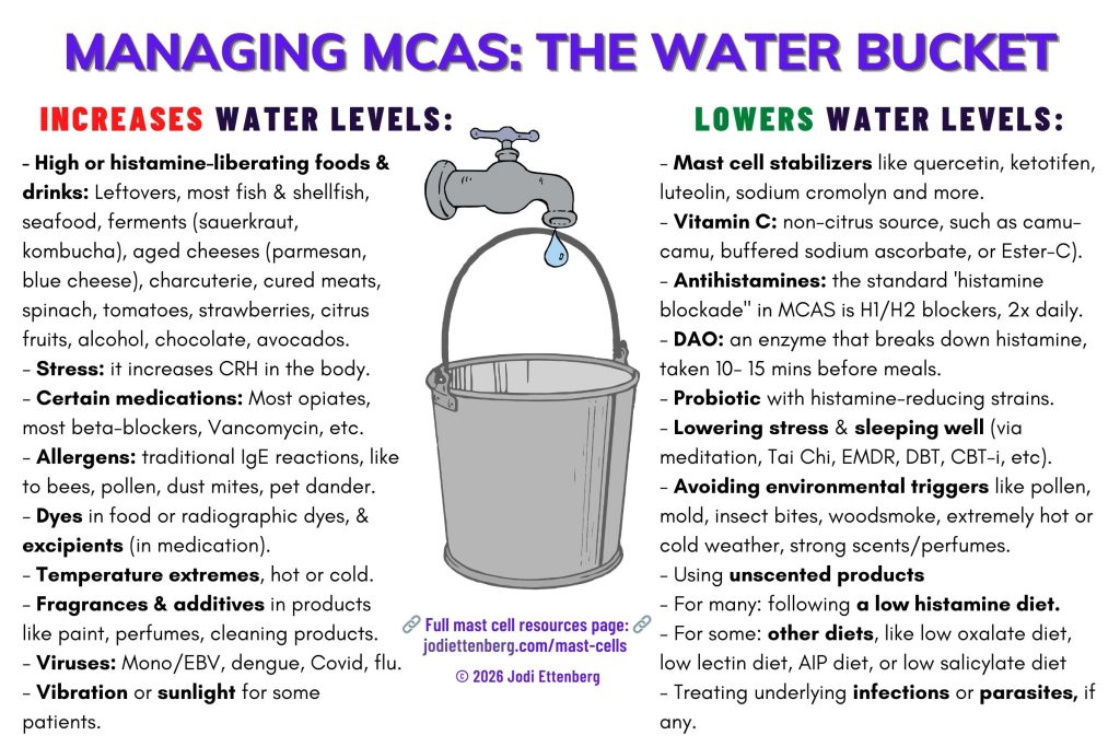
The “Bucket Analogy” in Mast Cell Activation Syndrome
With mast cell activation syndrome, exposure to triggers leads to reactions all over my body—sometimes culminating in anaphylaxis—but the severity of the reaction depends on a variety of factors all funnelling into my immune system.
Unlike with traditional allergies, there are times that exposure to mast cell triggers is slightly less impactful than others. It all depends on the amount of water in my bucket. Once the water in the bucket overflows, I stay in a prolonged mast cell flare. So my goal is to keep the water as low as possible, to prevent that from happening.
Here’s what I find raises and lowers the water for me in my “mast cell bucket“:
Sample script for friends and family
Here’s how I use the bucket analogy to describe what’s going on:
“Imagine a bucket where many things cause water to flow into it. Most people have a drain that works and is fully open in it. I have two problems: my drain is pretty clogged up, and I have a lot of water flowing into the bucket. When the bucket is super full, even a drop of extra water will cause the bucket to overflow.
Overflow can be gastrointestinal symptoms, itching, nausea, bone pain, [INSERT YOUR SYMPTOMS] and much more.
So my goal is to keep my water levels as low as possible. To do that, I go on a low histamine diet (helps less water flow into my bucket), I take mast cell stabilizers and antihistamines (which do the same), take DAO (an enzyme that breaks down histamine) with meals, which helps clear the drain a little bit.
But it takes constant calibration and also a calculus to try and make sure I keep the water low, and the drain as clear as possible. Keeping the bucket from overflowing also requires that I stay aware of so many little things that may go into the bucket. Environmental triggers, food, smells, and a lot of other little things that most people don’t need to pay attention to. I never needed to care about these things, but now only a small amount of exposure can lead to me being very sick for days.
So while I may appear over-cautious, or fussy, in reality it is what I need to do in order to stay as healthy as possible with this complicated condition“.
SYMPTOMS AND TRIGGERS OF MAST CELL ACTIVATION SYNDROME
What are the Symptoms of Mast Cell Activation Syndrome?
Because mast cells exist in so much of the body’s tissues and systems, when they degranulate and/or secrete mediators a large range of symptoms throughout the body may occur.
- Flushing of the face, neck, and chest
- Itching, +/- rash
- Hives, skin rashes — see the Mast Cell Society’s visual guide to skin lesions for more.
- Angioedema (swelling)
- Nasal itching and congestion
- Wheezing and shortness of breath
- Throat itching and swelling
- Headaches
- Alcohol-withdrawal headaches specifically, via the mast cell receptor MRGPRX2 (called Mrgprb2 in rodents). The receptor was long thought to be unique to mast cells, but it has since been found on basophils and eosinophils as well, and it was originally described in sensory neurons.
- Brain fog and cognitive dysfunction, accompanied with anxiety or depression
- Diarrhea, nausea, vomiting, abdominal pain, bloating, gastroesophageal reflux disease (GERD)
- Bone/muscle pain, osteosclerosis, osteopenia, osteoporosis
- Lightheadedness, syncope/fainting
- Rapid heart rate, chest pain
- Low blood pressure or high blood pressure at the onset of a reaction, depending on the patient; blood pressure instability
- Uterine cramps or bleeding
- Tinnitus / ear ringing (see the “other conditions” section below as well)
- Dermatographism (can write on your skin leaving a red welt where you traced something with a fingernail or blunt object; more here.)
- Edema (fluid accumulation in different parts of body)
- Decreased wound healing
- Interstitial cystitis
- Coagulation issues and blood disorders (Clots, deep vein thrombosis, easy bruising, heavy periods, nosebleeds and/or cuts that won’t seal up easily)

Possible effects of different mast cell mediators on the body
When mast cells degranulate and/or secrete mediators, these mediators can impact the body in different ways. While there are hundreds of mediators released by the mast cells, here are several of the most commonly cited ones, and their effect on the body:
Histamine: Flushing, itching, diarrhea, hypotension, panic attacks/anxiety
Leukotrienes: Shortness of breath
Prostaglandins: Flushing, bone pain, brain fog, cramping
Tryptase: Osteoporosis, skin lesions
Interleukins: Fatigue, weight loss, enlarged lymph nodes
Heparin: Osteoporosis, problems with clotting/ bleeding
Tumor Necrosis Factor-α: Fatigue, headaches, body aches
Symptoms of Mast Cell Activation Syndrome by Area of the Body
Some readers find it helpful to think of symptom clusters by area of the body, which can also make it easier to sequester symptom clusters by medical specialty. I will say that in my experience, few practitioners have been willing to explore mast cells as a cause for my symptoms. The only times that they’ve been considered is when labs were normal and my symptoms disappeared once I increased a mast cell stabilizer.
Still, the disease is systemic and it can be helpful to examine symptoms by area. When I note symptoms I get, it refers to times when my mast cells are not under control. These symptoms go away when things are stable for me.
Heart: Patients report palpitations, tachycardia, lightheadedness, out of breath feeling when active, chest pressure or pain, atherosclerosis, hypotension or hypertension, and more. Ventricular fibrillation, coronary artery disease, Kounis Syndrome and cardiac arrest can also occur in mast cell patients due to excessive activation of the mast cells.
GI System: Reported symptoms include abdominal pain, bloating after eating, nausea, constipation or diarrhea (or, as a friend says of her issues: the constant roller coaster between the two), gastroparesis, heartburn, irritable bowel syndrome. [Source]
Skin: Skin writing (dermatographism – my skin “lights up like a Christmas tree”, said my mast cell doctor), different rashes or red or brown spots that differ from the skin lesions in cutaneous mastocytosis, flushing (on the face and generally), stretch marks on the trunk or armpits, delayed wound healing (an issue for my spinal CSF leak!), ridges on the nails (also common in connective tissue disorders), hair loss, easy bruising, and itching. [See: “A Review of the Dermatologic Symptoms of Idiopathic Mast Cell Activation Syndrome”, here.]
Autonomic nervous system: While Postural Orthostatic Tachycardia Syndrome (POTS) is its own condition, there’s some evidence to suggest a connection with MCAS. Mast cells are localized close to blood vessels and peripheral nerves, and are also strategically positioned to modulate sympathetic activity and vascular tone. There are some cases of symptoms that doctors thought were hyperadrenergic POTS, but were actually MCAS —and the POTS-like symptoms resolved with stabilizers and antihistamines. See this case study. Of course it’s possible for patients to have both, but MCAS can affect the nervous system, and I felt this was worth mentioning.
Eyes, ears, nose, and throat: “Filmy” eyes are common, as well as irritation that is found in allergies (burning in the eyeballs), as well as sensitivity to bright lights, sun, and difficulties focusing vision. I get tics where my eyelids spasm when my mast cells are not well controlled. Tinnitus is very common (ringing in the ears), as well as sound sensitivity and occasionally reduction in hearing, mouth ulcers or canker sores (I get these frequently), burning pain on the tongue and mouth, geographic tongue, white patches on the tongue, problems with teeth and gums (patients report “crumbling teeth”, frequent cavities, and bleeding gums even though they take good care of their teeth and don’t eat much sugar), sore throat / red throat (I get this, alongside a hoarseness in my voice, especially upon waking when my mast cells are not controlled), post-nasal drip, sinus congestion. [More, via MastAttack]
Lungs/Respiratory: Pain or pressure in the respiratory tract, sporadic wheezing or coughing, difficulty breathing, and as with Ehlers-Danlos Syndrome, obstructive sleep apnea is common. Mast cells also contribute to pulmonary fibrosis, and this is an area of study presently.
Lymph nodes/Lymphatic: Enlarged lymph nodes, for me usually armpit or neck. Excessive mast cell activation can lead to lymphatic permeability and vascular remodelling, as well as immune cell trafficking through the lymphatic vessels, making the cross-interaction between mast cells and other areas of the body a topic of study presently especially as it relates to cancer. [See “Emerging Roles of Mast Cells in the Regulation of Lymphatic Immuno-Physiology”, here.]
Urinary and Gynecological: I hear a lot about uro-gyn symptoms from readers, and the symptoms can be very uncomfortable. Symptoms include increased urinary frequency, incomplete urination, flank pain/burning, “feeling like I have a UTI” (painful peeing, burning in the genital tract when not urinating, and more)— but testing is negative, and symptoms ebb when mast cell medication is taken. Interstitial cystitis diagnoses are common. Genital tract symptoms are less common in men, and in women include vulvodynia, vaginitis, and/or itching and redness (readers thought they had a yeast infection or bacterial vaginosis, but it went away with mast cell treatment), painful intercourse, and bleeding during or after intercourse. [See here and here for urinary issues; here for gynecological issues, and with more on mast cells affecting pregnancy and postpartum: here, with the full study here.]
Neurologic and Psychiatric: I remember reading a case study where a doctor reported a patient who said their “panic attacks went away when I started my antihistamines”. This was early on in my mast cell journey, and since then I am constantly amazed at how much mast cell degranulation affects mood, sleep, and more. Neurological symptoms include headache (often around temples; a “helmet” headache), dizziness, tingling, changing colour in hands and feet (for me, red and blue), burning in extremities, frequent nighttime waking but also inability to fall asleep, myoclonic jerks (jerking awake when falling asleep), night terrors, and a few of you write in to also report sleep paralysis. In addition, psychiatric symptoms that include anxiety (“physiological anxiety” is how some readers described it), panic attacks, depression, “histamine rage” aka irrational anger, brain fog, word finding problems and other cognitive dysfunction, [See here, with the full text of the study here, and a write up from Mast Attack here. Please also see the section on small fibre neuropathy]
Blood sugar, diabetes: High glucose levels, big fluctuations in glucose levels, diabetes mellitus, and more. Histamine affects insulin production, but the range of instability in patients using continuous blood glucose monitors is significant, so I hope we get more data as to why this happens, soon. [See here, here, and here (“observations from animal and human studies have suggested beneficial effects of treating diabetic patients with MC stabilizers“—but it’s not one size fits all)]
Cholesterol: It is common to have high cholesterol with mast cell activation syndrome. Mast cells are also involved in plaque (cholesterol) buildup in arteries, as noted in the heart section. [See here, here (includes liver abnormalities as well), and here (includes metabolic issues as well).]
Immune system generally: Lots and lots of reactivity (not always IgE allergies) and sensitivities to the environment (for me, a new pollen, scents, and fragrances issue), slow healing from colds or infections, and often the presence of autoimmune conditions/diseases.
What are Triggers for Mast Cell Activation?
Triggers for mast cell degranulation are all across the map, from food to activity to environmental exposure and trauma. It’s difficult to imagine just how such disparate and seemingly innocuous things (vibration?!) can degranulate mast cells or cause them to secrete mediators, but here we are.
The list below was created from TMS, The Canadian Mastocytosis Society, and blogs from around the web:
- Intense exercise
- Heat, cold or sudden temperature changes
- Sun/sunlight
- Fatigue
- Stress, trauma, loss, emotional pain, family problems
- Physical triggers, including pain, accidents, dental procedures, radiological dyes, surgeries, and other medical procedures. Both the Canadian and American Mastocytosis Societies recommend premedicating before any such procedures or dyes are administered. Radiation can also trigger mast cell issues. I had no problems with MRIs previously, but now have to premedicate even for non-contrast MRIs. For medical procedures, radiological procedures, dental surgeries, and more see this 2023 PDF guide to bring to the hospital with you that discusses mast cell disorders, plus stages of anaphylaxis and triggers. The ER protocol is often updated; see TMS’s resource page for more.
- Environmental (weather changes, pollution, pollen, pet dander, mold, gas leaks)
- Food or beverages higher in histamine. See the diet lists below under TREATING MCAS for more details. A big problem for people trying to get well is that many of the items on the “no” list for histamine are actually recommended enthusiastically for autoimmune diets, such as fermented foods/natural probiotics, spinach, and more. Unfortunately, these will make MCAS symptoms worse for many patients.
- Yeast
- Alcohol
- Dairy (especially fermented dairy like kefir, blue cheeses, or aged dairy like Parmesan cheese)
- Gluten
- Fermented foods (especially sauerkraut, kombucha, miso, kimchi, fish sauce, and soy sauce — anything that gives food a wonderful umami taste, basically)
- Cured and smoked meats and fish
- Shellfish
- Citrus fruits
- Vinegars
- Canned and processed foods (canned products tend to have higher levels of histamine than fresh ones).
- Overripe fruit and vegetables
- Leftovers — I can tolerate dinner food the next day at lunch but that’s it. (Usually, I will just freeze leftovers right away to later defrost to eat. This it stops the aging/histamine degradation process.)
- Berries, especially strawberries
- Spinach
- Chocolate other than pure dark chocolate
- Tomatoes
- Some food additives (see here for more)
- A surprising number of medications. The ‘avoid’ list includes some of the below. A longer list is here, and here. The latter list also includes medication that may work to calm mast cells.
- Angiotensin converting enzyme inhibitors (ACE inhibitors) used to treat high blood pressure/hypertension can also increase bradykinin levels and therefore stimulate activation in mast cells
- Amphetamines
- Aspirin
- Beta Blockers – many readers and studies note that beta blockers (for tachycardia, POTS, and more) can raise the floor on mast cell issues, making mast cells more likely to degranulate. This in turn can cause issues if a patient also needs epinephrine during anaphylaxis.
- Dextromethorphan (cough suppressant)
- Dipyridamole (Persantine)
- Fungal infection drugs
- Local anesthetics: some local anesthetics like benzocaine, tetracaine, and chloroprocaine, can trigger mast cell activation. Lidocaine or Carbocaine seem the best tolerated, but ask if there are preservative-free versions available.
- Neuromuscular blocking agents (all), such as Decamethonium, Gallamine triethiodide
- NSAIDs (Non-steroidal anti-inflammatories like Advil ,Motrin, etc.): these can sometimes help patients, but in many they cause excess degranulation. You’ll have to figure out which category you fall into.
- Most opiates (Codeine, Morphine, Percocet/Oxycodone, etc.): from what I’ve read, fentanyl, Dilaudid, and occasionally Tramadol are best tolerated.
- Thiamine hydrochloride (A form of vitamin B1)
- Tolazoline hydrochloride
- Trimethaphan
- Vancomycin, often used with C. difficile
- Contrast dyes used in MRIs, CTs etc, whether iodine contrast agents or gadolinium. Some patients report gadolinium to be better tolerated, but others say it makes them feel sicker.
- Mechanical irritation (rubbing/chafing) or friction
- Vibration (for me, this included the MRI machine! I got a 3T MRI and had to take extra antihistamines as my body was jerking/moving despite my trying to stay still. This never happened before 2020, and I don’t fear or have claustrophobia or other concerns with MRIs. This new reaction truly took me by surprise.)
- Natural odours (strong natural scents like essential oils, environmental smells)
- Strong odours (cleaning products, for example, or perfumes)
- Infections (viral, bacterial or fungal infections can make MCAS worse and should be addressed carefully with help of your doctor – see below for how that works, including the image I included)
- Venom (bees, wasps, spiders, snakes, etc.)
- Diesel and other fuel (or more specifically the smell thereof)
Viruses like Covid, dengue fever and EBV can sometimes trigger mast cell activation
Mast cells are involved in the body’s normal antiviral response, but in some circumstances, prolonged immune activation or interactions with other parts of the immune system may contribute to sustained mast cell activation.
For example, what kicked everything off for me as an adult was getting dengue fever from a mosquito on my travels. There are quite a few studies looking at the role of mast cells in dengue infections, including one that notes:
Immune cells called mast cells can hinder rather than help the body’s response to dengue virus, which suggests that mast cell products could be used as biomarkers to identify severe forms of the disease.
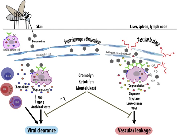
As the caption to the (ADORABLE) image notes, mast cells can be a double-edged sword in viruses, because we want them to do their job and clear out a virus, but when they are dysfunctional, they overdo it. And in non-MCAS patients, trying to tamp down on the response may, the study notes, prevent them from doing their job in clearing the virus altogether.
Studies relating to influenza also note that tamping down on mast cell activation can be beneficial. A study “Mast Cell-Induced Lung Injury in Mice Infected with H5N1 Influenza Virus” notes,
A combination of ketotifen and the neuraminidase inhibitor oseltamivir protected 100% of the mice from death postinfection. In conclusion, our data suggest that mast cells play a crucial role in the early stages of H5N1 influenza virus infection and provide a new approach to combat highly pathogenic influenza virus infection.
Taken together, these studies suggest that viral infections such as Covid, dengue and influenza can activate mast cells, and that mast-cell activation can have both protective and potentially harmful effects. Whether a particular person’s mast-cell response contributes to prolonged symptoms after a viral infection is much less well established, though.
For people who already have mast-cell problems, it is plausible that an infection could exacerbate those symptoms, but this has not been established as a general mechanism.
In 2024, the American Centers for Disease Control and Prevention (CDC) updated their post-viral illness page to list out viruses that can lead to long-term symptoms in the body, and included Covid, dengue fever, and EBV as some of the viruses known to do so.
DIAGNOSIS OF MAST CELL ACTIVATION SYNDROME
As I wrote above, diagnosis of mast cell activation syndrome (or a sister disorder like mastocytosis or HαT) is often delayed due to lack of awareness about the nature of the condition and/or some practitioners testing only a narrow selection of mediators or only testing IgE allergies.
In addition, the lab tests required to diagnose MCAS must be handled with great care and kept chilled. Specimens for urine testing also need to be kept cold at all times. The result is that several rounds of testing may be needed to confirm the lab results — which can be quite frustrating.
A differential diagnosis analysis is usually applied by the practitioner to rule out other inflammatory immune conditions or mimics of a mast cell disorder. Symptoms are so bio-individual, too, that it’s a lot harder to pin down a diagnosis than (say) a condition where it’s a simple gene test. The labs below test for some mast cell mediators to determine their ranges, but experts have noted there are hundreds of mediators released by the mast cells in total. Thus far, only a handful of them have lab tests to measure their values.
For those in Canada, doctors often don’t run (or can’t order) many mediator tests as they are expensive and not covered by provincial healthcare. Some private mast cell practitioners will cover them, as will mast cell specialists in the United States.
What labs, urine testing, and DNA tests are used to test for MCAS?
Standard testing for MCAS includes both urine and serum (blood) tests. It can sometimes involve biopsies of the GI tract or colon. As stated earlier, samples can degrade really quickly at room temperature, and must be handled properly or else they will become corrupt. The mast cell doctor I saw said that laboratory or patient handling mistakes account for a lot of the false negative testing he sees, and he only works with specific labs that he knows he can trust to properly chill the samples.
Here were the tests the specialist ordered for me in order to assess whether or not I had mast cell activation syndrome:
Specific blood tests for MCAS that were ordered for me:
- Histamine, blood
- Histamine, plasma (chilled)
- PGD2, plasma (chilled)
- Comprehensive Metabolic Panel
- PT + PTT
- Plasma heparin level Anti-Xa (anti-Xa for unfractionated heparin, chilled)
- Tryptase, Serum
- Chronic Urticaria Index
- Anti-IgE antibodies
- Chromogranin-A
Specific urine tests for MCAS
- Chilled Histamine, 24hr Urine
- Histamine, Random Urine (then chilled)
- Random urinary prostaglandin D2 (then chilled)
- Chilled 24-hour urinary prostaglandin D2
- Random urinary 2,3-dinor-11-beta-prostaglandin-F2-alpha (then chilled)
- Chilled 24-hour urinary 2,3-dinor-11-beta-prostaglandin-F2-alpha
- Random urinary N-methylhistamine (then chilled)
- Chilled 24-hour urinary N-methylhistamine
- Random urinary leukotriene E4 (then chilled)
- Chilled 24-hour urinary leukotriene E4
DNA Tests
- Autoinflammatory Syndromes Genotypings Panel (originally from Invitae, now run by Labcorp Genetics as Invitae filed for bankruptcy in 2024 and Labcorp acquired its testing assets)
Consensus about mast cell activation syndrome is lacking in the field
Among the mast cell experts, there are currently two ‘consensus guidelines’ related to diagnosis, both published within the field but representing a significant divergence in opinion among experts. The guidelines address not only who should be diagnosed with the condition, but also how to manage the care of patients who do not meet the diagnostic criteria. Both sets of criteria require that the patient has symptoms consistent with chronic aberrant mast cell activation and that no other disease could be identified that accounts for the full range and duration of what the patient is experiencing. They also require that those signs and symptoms involve at least two organ systems. The Consensus 1 framework places substantially more emphasis on demonstrating a rise in tryptase, whereas Consensus 2 does not require tryptase elevation.
The Consensus 1 group consists of Drs. Valent, Akin, and others, and their consensus criteria are here.
Consensus 1 is the framework endorsed by the American Academy of Allergy, Asthma & Immunology, and requires all of the following:
- Symptoms typical of mast cell activation that are severe, episodic and recurrent, and that involve at least two organ systems (often discussed as recurrent anaphylaxis).
- Objective laboratory evidence that mast cell mediators rose during a flare. For serum total tryptase, this means a rise of 20% above your own baseline tryptase, plus 2 ng/mL, measured during or shortly after an episode. Where that cannot be demonstrated, elevations in prostaglandin D2 or its 11-β-PGF2α metabolite, or in urinary N-methylhistamine, can be accepted instead.
- Improvement on medications that block mast cell activation or its mediators, meaning at least a partial response to antihistamines and/or or sodium cromolyn.
The Consensus 2 group consists of Drs. Afrin, Molderings, and others, with their criteria here. Consensus 2 recognizes a wider range of symptoms than Consensus 1, all of them noted as consistent with the known effects of mast cell mediators. Consensus 2 requires chronic or recurrent multisystem problems, generally inflammatory and often with allergic or dystrophic features, affecting at least two organ systems. Symptoms are assessed together rather than individually.
It also permits broader mediator testing to be relevant to diagnosis: tryptase, chromogranin A, heparin, prostaglandin D2, histamine, N-methylhistamine, 11-β-PGF2α, and leukotriene E4. Rather than requiring a rise measured against the patient’s own baseline like Consensus 1, Consensus 2 criteria discuss a level above the ordinary reference range, read in the context of otherwise unexplained chronic multisystem illness. For treatment response, Consensus 2 notes that improvement on mast cell-targeted therapy can count as a minor criterion, but does not require it. The authors object to making it mandatory because they are unaware of other diseases whose diagnostic criteria require a therapeutic response, and because requiring it may lead to patients being excluded simply because they did not have success with a short list of drugs.
Both groups published updates on their positions in 2026. The Consensus 1 group published in January in a commentary review entitled “Diagnosis and Management of Patients With Mast Cell Activation Syndromes: Status 2026“. They argue that referrals to specialist centres have risen substantially over the past fifteen years, and attribute this to a combination of increased awareness, the ordinary prevalence of IgE-mediated allergy, and patients and clinicians arriving at MCAS through unverified information online. Their review reiterates that tryptase remains the marker they consider most validated for the condition.
The Consensus 2 group published the six-year review article in June 2026 entitled “Progress in mast cell activation syndrome: the global consensus-2 diagnostic criteria at six years“. They argue that their broader criteria have allowed diagnosis and treatment options to be afforded to patients who were previously labelled with inflammatory syndromes or misdiagnosed as somatization. They also note that the more restrictive criteria are underdiagnosing a treatable condition, and that the overdiagnosis predicted when Consensus 2 was published in 2020 has not occurred.
The concern on the Consensus 1 side is that a diagnosis applied too readily can interfere with searching for a different cause. Dr. Akin has noted that questioning whether a patient meets Consensus 1 MCAS criteria does not undermine the patient’s symptoms; those symptoms are still real. Unfortunately, patients write in frequently to share that they were told that their symptoms were psychosomatic, and instead of exclusion leading to more investigation, faced a diagnostic dead end.
In a September 2026 article, “Mast Cell Activation Syndrome Diagnosis Divides Experts as Patients Look for Answers” on Medscape, mast cell expert Dr. Theoharides notes the two risks clinicians ultimately need to navigate between that reflect the dispute in criteria, too: either attributing too many symptoms to mast cell activation, or overlooking the condition when it may be contributing to a patient’s illness.
Tryptase in patients with suspected mast cell activation syndrome
Since MCAS is a recently defined condition — the first proposed diagnostic criteria date from 2010 and 2011 — some allergists or immunologists will test tryptase and, if it’s negative, say it’s not MCAS. As noted above, there is some dispute among MCAS specialists as to whether tryptase must be elevated in order to be diagnosed with the condition. Many mast cell experts, even those who include tryptase in the requirements for diagnosis, do test other mediators, however. Tryptase testing alone is not sufficient to rule out MCAS.
In a podcast about Consensus 2, Dr. Afrin responded to comments about why tryptase should not be a reliable / gold standard marker for diagnosis of mast cell activation syndrome, as follows:
[I]n patients who have the activation syndrome, typically tryptase is not elevated at all. In fact, the published research suggests the tryptase is elevated in only about 15% of MCAS patients. And even in those 15%, we now understand there’s something else going on besides mast cell activations actually driving the modest elevation of tryptase. […] Im the 85% or so of MCAS patients who don’t have an elevated tryptase, it’s instead these other markers of mast cell activation that you have to go looking for to prove that.
However, as mentioned above, tryptase is elevated in both mastocytosis (but not all cases of it) and in HαT.
If your tryptase is elevated, you may want to consider testing for HαT, Hereditary Alpha Tryptasemia, which is newer in the mast cell disorders world. It is an easier condition to rule in or out, because it requires only a copy-number test of the TPSAB1 gene, rather than the finicky mediator testing MCAS requires. Gene by Gene offers a saliva-based test that can be ordered online. ARUP Laboratories also runs a TPSAB1 copy number analysis test. HαT affects roughly 4 to 6% of the general population and is a common cause of an elevated baseline serum tryptase.
In addition, you may qualify for a noninvasive KIT D816V blood test for systemic mastocytosis, which can be tested via Labcorp’s Blueprint biomarker testing program. As systemic mastocytosis is characterized by neoplastic proliferation of mast cells driven by the KIT D816V mutation in more than 90% of the condition’s cases, this biomarker is an important test. Note, though, that a negative blood KIT D816V test does not rule out systemic mastocytosis, and a positive result does not necessarily eliminate the need for bone marrow evaluation. The blood test may help determine whether further evaluation, including bone marrow biopsy, is warranted. At the time of publication, this test is provided by Labcorp Oncology for eligible patients at no-charge through a sponsored testing program from Blueprint Medicines.
Notes about mast cell testing, HαT, & mastocytosis: please read before testing!
Going off medication before testing
- It is not a given that you will be asked to go off your antihistamines for testing. In Canada, some doctors do require this. In the United States, most mast cell specialists do not. Do not stop medications on your own before testing; whether a particular medication should be withheld depends on the test and the clinical situation, so follow the ordering clinician/laboratory’s instructions.
- Some doctors do require you to get off mast cell stabilizers for testing. I was instructed to go off quercetin, PEA, Vitamin E, and the other stabilizers listed below one week prior to testing, as well as anything that affected inflammation.
- If you take NSAIDs like Advil, you are generally asked to stop 5 days prior to your testing.
- If you take PPIs (proton pump inhibitors) you are generally asked to stop 5 days prior to your testing.
IgG vs. IgE reactions
- There’s often confusion about testing for food allergies or allergies using IgG and IgE testing. They are related to immune responses that can involve mast cells, but not an accurate test for mast cell activation.
- Mast cells have receptors for several types of immunoglobulins, including IgE and some IgG subclasses. IgE is the antibody class most strongly associated with classic immediate allergic reactions, while IgG antibodies have different and more complex roles in immune regulation and inflammation.
- IgE allergens can lead to mast cell degranulation. But IgE testing (traditionally done by an allergist) is not a criterion for a diagnosis of MCAS. You can have no IgE allergies and still have MCAS.
- Many people with mast cell disorders have physical urticarias such as dermatographism, where light scratching causes raised, red welts. I can easily write my name out on my skin and it’ll glow puffy and red for a long time. Dermatographism can make skin-prick testing more difficult to interpret, but neither skin-prick testing nor IgE testing is a test for MCAS.
- The presence of IgG antibodies to a food does not, by itself, lead to the conclusion that the food tested is causing symptoms, even though commercial food IgG panels sometimes are marketed as a tool to find “food sensitivities” or foods that are causing inflammation. There is currently no evidence that these tests can diagnose food allergy, food intolerance, or MCAS. Major allergy organizations recommend against using them for these purposes. A positive food IgG result generally means that the immune system has been exposed to that food, and has produced antibodies to it. It does not mean that the food is triggering mast cell activation necessarily. IgG-mediated immune mechanisms are relevant to mast cells, but a consumer IgG panel won’t tell you what is causing your specific symptoms.
- IgE reactions, in contrast, are often sharp and immediate. For example, being stung by a wasp if we are allergic to wasp venom. IgE reactions consist of a fast-paced degranulation response by the sentinels of the immune system to try and protect from something significant. While not part of MCAS testing per se, they can be useful.
BRIEF SUMMARY OF HOW I GOT MY MAST CELLS UNDER CONTROL
The level of pain I experienced with a spinal CSF leak and the (unknown to me at the time) mast cell dysfunction was debilitating. It got to a point where I simply sobbed in bed all day long, my body feeling like it was on fire and nothing could stop the pain. When my research led me to suspect a mast cell disorder, I implemented the following steps. They sharply reduced my pain levels while I waited for specialist testing.
** This is just what has worked for me so far and is not medical advice. **
1) I identified triggers and removed them from my environment, like strongly-scented home cleaning products, non-natural cosmetic products or shampoos or face washes (see below for what brands I use), and more. This includes getting air purifiers for my apartment, to help rid the air of triggers I may react to. In my part of Canada, fireplaces or wood smoke heating is common in the winter; I also had to make sure I could turn off my air exchange and limit incoming air during those months, or else I got very sick. Some options: a Levoit Air Purifier (this is their larger room model, but they have smaller sizes that are cheaper) is one I recommend to many as it is less expensive. For a heftier option, there is Austin Air. They are also more expensive, but note that filters need to be changed only every 5 years. For Canadians, there is AtmosC, who have smaller and larger sizes, and a 9-phase filtering system that includes a HEPA filter.
2) I went on a strict low histamine diet for two weeks, removing things like alcohol, cold cuts and smoked/cured meats, canned fish, anything pickled or fermented, aged cheese, overripe produce, gluten (already done because I’m celiac), citrus fruit, spinach, yeast, soy, and tomatoes. I also stopped eating leftovers more than one day old, freezing anything I cooked after making it for future consumption. See the diet list below for the full list of items to remove. As I identified a list of what were my worst triggers (for me these include tomatoes, spinach, aged cheeses and charcuterie, and more), I slowly re-added in some of the medium histamine foods as able. Ketotifen was needed for me to be able to do so.
3) I added natural mast cell stabilizers like quercetin and PEA. See the “what I take” list below for what I used.
4) I made some additional lifestyle changes: a) starting DAO, the enzyme listed below, 15-30 minutes prior to eating higher-histamine meals, and b) reducing stress by continuing with a meditation practice. Lowering stress can reduce levels of corticotropin-releasing hormone (CRH), a mast cell degranulator. Not an easy task in today’s world, I know. Or when your body feels like it’s running away with the prize.
5) I started trialling different H1 antihistamines to see which of the available over-the-counter options helped me the most. If an H1 didn’t work or make a big difference after two weeks, I stopped it and tried a different one. The standard dosing regimen for MCAS is that H1s/H2s are taken every 12 hours to create a “histamine blockade”. This exceeds the label instructions for H1s, but is normal for MCAS.
6) Once I had an H1 blocker on board that worked for me (it was levocetirizine that worked best; other H1s caused side effects, or did not manage my symptoms as well as levocetirizine), I started the same trials with H2 antihistamines. I found that ranitidine (MAR-Ranitidine in Canada, formerly OTC as Zantac) at low doses, was the one that worked best for me, and I still use it many years later. This was pulled from the market in the USA and Canada a few years ago over concerns about NDMA contamination. The FDA approved a reformulated ranitidine tablet in November 2025, so it is back on the US market by prescription only, not as OTC Zantac. The new labelling carries storage instructions that include discarding unused tablets 90 days after opening. In Canada it was available and batch tested throughout (I’ve taken it without any gaps). Since ranitidine was pulled for some time, many patients use famotidine (Pepcid). These H2s are taken as noted above, with the H1 every 12 hours — again, not as directed on the packaging. There are H2 receptors in the GI tract, but also in the brain and smooth muscle, and heart. Blocking H2 receptors as an MCAS patient helps with a variety of symptoms and can even help certain types of POTS.
7) To ensure some of my mast cell dysfunction wasn’t being fuelled by parasites or viruses after a decade of eating my way around the world, I did a stool test and blood test. They did not turn up any additional surprises.
For each of these steps, it was important to go low and slow as I onboarded new supplements or medication. It’s very difficult and can be frustrating to identify triggers, and one of the most difficult is excipients or fillers in supplements, foods, or medications. I now have a long chart where I write in the supplements or medication that work for me, and their excipients or fillers as well, because mast cell experts have made clear that excipients or fillers in medication are often what mast cell patients react to and not necessarily the medication itself. Here is more on excipients: Recognition and Management of Medication Excipient Reactivity in Patients With Mast Cell Activation Syndrome, a 2019 review article.
Unfortunately, a 2024 study revealed that the main place patients go to look at excipients in North America, the DailyMed database, is not as accurate as we thought. The study found that internal inconsistencies of excipients occurred in 39% (!) of the formulations examined. So it’s best to go straight to the product monograph, if you can.
Briefly, some of the excipients readers have reported back on that give them trouble are:
- magnesium stearate
- polyethylene glycol
- coatings on ‘extended release’ or ‘enteric’ pills, including shellac or dyes like FD&C Red #40, FD&C Red #3, or FD&C Blue #2. (FD&C Red #3 is close to eliminated in the USA; the FDA revoked its authorisation in January 2025, with a deadline of January 2027 for food and January 2028 for ingested drugs.)
- povidone
- alcohol
It wasn’t fun or easy, but with no access to knowledgeable medical care, it really helped me stay afloat until I could get to a mast cell specialist.
TREATING MAST CELL ACTIVATION NATURALLY
Eliminating Triggers
Reduction of triggers includes whatever triggers you from the long list I shared earlier. This means removing not only environmental triggers like shampoos and soaps, but for many patients, eating a lower histamine diet to potentially lower circulating levels of histamine in the body.
Note that for many vibration is a trigger, so car rides, electric toothbrushes, and more can degranulate mast cells for those patients. Please see the list of triggers above for more help in narrowing down what may need to be tested in your own case.
Low Histamine Diet
Few mast cell patients I’ve spoken with eat a normal diet. As a former food/travel writer, this part is hard. I joke that I’m the worst food writer ever, but the truth is after wrapping my head around celiac disease 20 years ago, cutting things out strictly and systematically is not new to me. I just didn’t ever think I’d have to cut out so much!
Being on a low histamine diet can help reduce the overall burden on your body because it reduces the amount of histamine floating around. For non-MCAS patients, it can also sometimes help itching or asthma.
Some doctors do not recommend a low histamine diet, or maintain that it is not helpful overall for the condition. They prefer to rely on medication and/or supplementation to best control symptoms, since histamine is but one mediator released by the mast cells. A 2021 study reviewed the food lists used in low histamine diets and found that high histamine content could account for excluding only 32% of the foods on them. It also found wide disagreement between lists, other than fermented foods; they were the only category every list excluded. The paper also noted that some foods appeared on these lists because they were thought to liberate histamine or interfere with DAO, rather than because they contained high levels of histamine.
That said, at a 2022 conference on mast cell activation syndrome that I attended, several practitioners specializing in mast cell activation (including a gastroenterologist) noted that it’s a must. It does appear that more doctors are seeing clinically that a low histamine diet can help symptoms. In my case, it has proved to be crucial. Without significantly limiting the food I take in, I cannot stay on top of my reactions and symptoms. My subsequent lab testing also revealed a very high level of circulating histamine — my physician said it was among the highest they’d ever seen in thousands of patients — so I can see why it helps me.
Also, why is my immune system an overachiever?!
While my mast cells do release far more than histamine when they are agitated, I’ve seen big strides in my day to day baseline by removing certain foods from my diet.
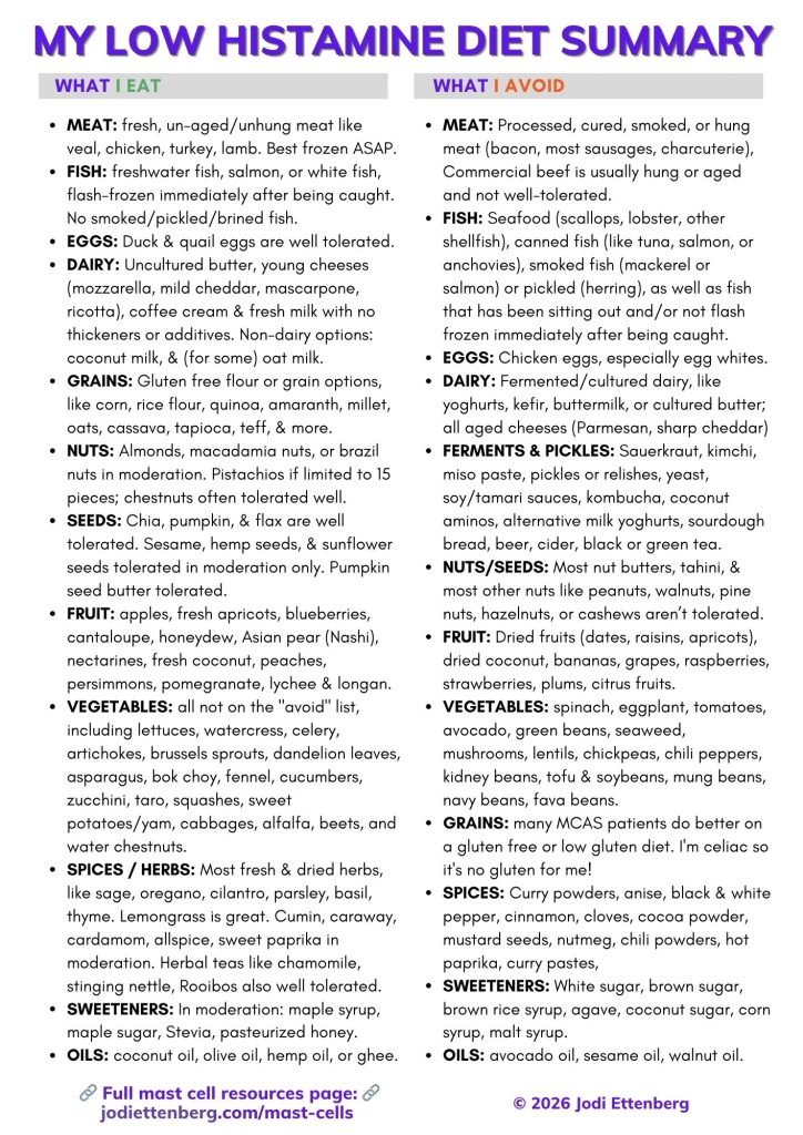
What to eat or avoid on a low histamine diet
As I keep saying in this post: everyone’s body is different. That said, there are some commonalities where certain foods or food products are high in histamine, or histamine liberators, and the bulk of the food lists out there related to the condition recommend caution.
I made a chart to break it down more easily. The chart synthesizes the many food lists out there and includes the many years of community feedback from patients, as well as my own dietary changes. (Right click to view in a separate tab and download the image). Note that since starting a microdose of a GLP-1, tirzepatide, I have been able to re-add many foods that I could not previously tolerate with MCAS, such as red grapefruit, plain yoghurt, and more.
Foods you can eat on a low histamine diet —
- Many of us do great on meat that is fresh and unhung/not processed or aged. This includes lamb, chicken, veal, rabbit, turkey, and more. Best practices are to try and find products that are butchered and immediately frozen or vacuum sealed (preferably both). Ground meat is a tough one because it’s higher in histamine. I’ve managed fine on meat that is immediately frozen after being ground up. In general, you want to minimize exposure to air so meat that is vacuum sealed and fresh will be a safer bet than in loose packaging. Many products are vacuum sealed these days, so there is more to choose from.
- Fish that has been flash frozen after being caught, and not pickled/brined. I can tolerate salmon sashimi if purchased from frozen (sushi grade).
- Duck and quail eggs, though some people can tolerate chicken egg yolks.
- Dairy that is safe for us includes regular (uncultured) butter, young cheeses like mozzarella, mild/young cheddar, coffee cream, fresh milk (though not everyone can tolerate), mascarpone and ricotta cheeses. Some people can also do cottage cheese or cream cheese; since adding Ketotifen, I am one them.
- Gluten free flour or grain options, like corn, rice flour, quinoa (high in oxalates though), amaranth, millet, oats, cassava, and more.
- For nuts, I do well with the occasional almond, Brazil nut, or cashew, and I tolerate macadamia nuts and pistachios just fine. For seeds, sesame seeds in moderation, chia seeds, flax seeds or ground flax, hemp seeds, and occasionally pumpkin or sunflower seeds. I also frequently roast the seeds from any squash I cook, which are always so tasty!
- Vegetables: basically, everything EXCEPT the things I list in the “avoid” section. In particular, people do great with lettuce, watercress, broccolini or tenderstem broccoli, chestnuts, celery, artichokes, brussels sprouts, dandelion leaves, asparagus, bok choy (I eat this a lot!), fennel, cucumbers, zucchini, taro, red cabbage, and more. Some people do struggle with arugula, however. Also great are squashes like acorn and butternut squash.
- Fruit: apples, apricots, blueberries (in smaller doses), cantaloupe, honeydew, Asian pear (Nashi pear), nectarines, peaches, persimmons, pomegranate, and lychee and longan are all low in histamine and delicious.
- Most fresh herbs and several spices, including cilantro, oregano, sage, parsley, basil, allspice (I use it in the place of cinnamon, which is high histamine), limited cumin, and cardamom. Herbal teas are not fermented, and I love chamomile tea, stinging nettle tea, Rooibos, and more. Lemongrass also ok to season dishes! I use sumac as a spice to give a bit of acid to foods, since I cannot tolerate citrus, including lemon or lime. Cooking with lime leaf, however, is tolerated — and delicious.
- I use maple syrup to sweeten baked goods, and tolerate Stevia and pasteurized (not raw) honey.
When cooking:
- Many dietitians and mast cell posts will say to avoid cooking at high heats, including charring and/or BBQing. At the beginning, I did boil instead of roast food, but as time went on and I could tolerate more foods once I found a polypharmacy approach that worked for me, I can now eat foods that are cooked at higher heats. Cooking oils that I use safely are coconut oil, hemp oil, olive oil, and ghee.
- Leftovers are an issue, because the longer a food product sits there, the more histamine rises. This also includes overripe fruit or veggies. Histamine in meat rises more quickly than vegetables; I can have leftovers the next day for meat, but two days when it comes to vegetables only. Rinsing your food off can sometimes help reduce surface histamine, say some experts. YMMV.
High histamine foods to avoid —
- Processed or cured meat, and beef: Meat that is smoked, cured (like bacon or charcuterie or sausages), or processed (sliced sandwich meat, hot dogs), and beef (as it is usually hung prior to being available for sale) are all high in histamine. If eating game meat, be sure to ask if it’s been aged or hung before it goes up for sale. Ground meat is also higher in histamine, and is best avoided unless it is ground and then immediately frozen, as per above.
- Most fish, especially seafood (scallops, lobster, other shellfish), canned fish (tuna, salmon, anchovies), fish that has been smoked (mackerel, smoked salmon), and fish that has been sitting in the fridge / store for some time.
- Some dairy: dairy that has been fermented (yoghurts, kefir, buttermilk, cultured butter), and aged cheeses like sharp cheddar, Parmesan, Gruyère, and more.
- Chicken eggs, especially egg whites, are high in histamine. As mentioned above, some patients can tolerate chicken egg yolks. Personally, I do best with duck eggs.
- Ferments: many diets that are espoused as “healthy” these days call for much spinach and ferments. These are histamine bombs. For ferments, we’re talking things like sauerkraut, kimchi, miso paste, coconut aminos (often better tolerated than soy sauce, though still fermented!), yeast, tamari and soy sauce, kombucha, and coconut milk / dairy alternative yoghurts. It also includes breads that are fermented, like sourdough, and beer and cider, and black and green tea, which are oxidized. Fermented teas like Pu-Erh tea are also off limits. It may be worth a test to see if you react; sadly I do.
- Pickled products: pickles! I love pickles. Sadly, these are now off limits and make me quite sick. Also add relishes, pickled onions, olives, and pickled carrots / cucumbers often found in Vietnamese food.
- Some vegetables: the biggest culprits are spinach, eggplant, tomatoes, avocado, green beans, mushrooms, seaweed/algae, chili peppers of all varieties, and for many people pumpkin, broccoli, and Swiss chard can also cause issues.
- Some fruit, especially dried fruit like dates, raisins, desiccated coconut, dried apples. As well as fresh fruits like bananas, grapes, strawberries, cherries, raspberries, and citrus fruits. I tolerate mango, but many do not. I can’t do grapes, but many others can. YMMV. Watermelon is also a hit or miss depending on the patient.
- Most legumes and pulses, including soybeans (and edamame), chickpeas, lentils, black or red kidney beans, fava beans, broad beans, and more. Tofu also is a trigger for most patients.
- Most nuts, seeds, and nut butters, especially peanuts, walnuts, tahini, and pine nuts.
- Grains: as a celiac I’ve avoided wheat at all costs for a long time, but many patients do find a gluten free diet helps their symptoms. This is a personal choice, but either way there are many great non-wheat options around these days. Buckwheat is also higher in histamine, despite being gluten free.
- Some spices, like black and white pepper, spicier curry powder blends, anise, cinnamon, cloves, cocoa powder (also high in oxalate), mustard seed, nutmeg, chili powders, and hot paprika.
- Oils: sesame oil, walnut oil, avocado oil.
- Sweeteners: white sugar, brown sugar, raw honey, agave, malt syrup, coconut sugar, brown rice syrup.
- Alcohol is high in histamine, especially fermented alcohol, but if you’re going to imbibe, the ‘safest’ option will usually be clear, distilled alcohols like vodkas or gins. I do find if I have white wine and use a “wine wand” I can have the occasional treat.
For me, a low histamine, medium oxalate diet seems to be the best baseline food plan, with occasional treats. I use a combination of the SIGHI list with Alison Vickery’s low histamine diet PDF:
- Low Histamine: Swiss SIGHI lists. Elimination diet recommendations here. Food lists for histamine: a long PDF of graded histamine levels in foods/additives, etc here.
- Low Histamine: Alison Vickery. Here is her DIET PDF. She uses a functional approach that combines naturopathic and allopathic medicine, and cites her sources fully. Of all the low histamine diet lists, this one has most matched how my body reacts to different foods.
Other diets that may help in stabilizing mast cells
In addition to low histamine, there are many patients who find extra relief using a low oxalate diet, especially for patients with pelvic inflammation or interstitial cystitis. A smaller subset of patients say they benefit from low FODMAP in addition to low histamine.
Some other “beyond low histamine” diet plans from around the web:
- Some people with Ehlers Danlos variations or auto-immune issues such as Crohn’s or ulcerative colitis also have trouble digesting foods beyond the low histamine varieties. See this page about those additional restrictions, including a low histamine diet suggestion list for people who need to be mindful of other categories.
- These categories include lectins, oxalates, salicylates, sulfur, and FODMAPs, which — depending on the body — can affect MCAS as well. Note that if oxalates are an issue, higher doses of vitamin C will be a problem. It is important to keep track of data related to food and supplements to ensure you can pin down triggers.
- Lectins are proteins found in some plants, and preliminary research suggests that they may activate mast cells also. For a low-lectin diet list, see here.
- Vitamin C is metabolised into oxalate in the body, so higher raise oxalate levels. While otherwise helpful for mast cell stabilization and for collagen synthesis, high dose vitamin C is something I’ve had to avoid in order lower oxalate intake. It took some time to realize that oxalates were worsningsymptoms for me. There are many oxalate food lists out there, but you can start here.
- FODMAPs: a low FODMAP diet can also reduce circulating histamine.
- From study: “Controversies and Recent Developments of the Low-FODMAP Diet” – “Mechanistically, dietary FODMAPs have very limited effects on the consistency of bowel actions but seem to suppress the release of histamine”
- And from the study FODMAPs alter symptoms and the metabolome of patients with IBS: a randomised controlled trial.“Histamine, a measure of immune activation, was reduced eightfold in the low FODMAP group (p<0.05)”
- NOTE: would need low histamine AND low FODMAP for this to work, as a few of the low FODMAP foods are high in histamine.
Apps that can help you manage low histamine and other diets
- Baliza is a German company that has developed several apps that are useful for the low histamine diet, which are updated as new data and studies come out:
- Their app Food Intolerances is very thorough and allows you to sort by different food intolerances or allergies. Includes measurements for histamine, fructose, sorbitol, gluten, lactose, and FODMAPs.
Apple here; Play store here. - Their app called OxiPur: measures foods by oxalate and soluble oxalate, searchable, sortable by food category, and includes some measurements related to oxalate content, like calcium in each food. Apple only, here.
- Their app Food Intolerances is very thorough and allows you to sort by different food intolerances or allergies. Includes measurements for histamine, fructose, sorbitol, gluten, lactose, and FODMAPs.
- Fig food scanner has a low FODMAP diet option, as well as other tweaks, and you can add foods you react to or ones you can’t tolerate. It lets you scan products from the stores, so it’s best for American readers as it’s based there. For Android here, or iOS here.
Supplements for Mast Cell Activation Syndrome
(Studies supporting why I take each of these products are below.)
- Seeking Health DAO: DAO is an enzyme naturally produced in the body that helps break down histamine from food intake. Some people have genetic mutations that lower their DAO production, some people eat diets rich in products that lower DAO, and some people just need the extra DAO to help them mitigate the effects of high levels of circulating histamine. There are two products on the market that are trustworthy for DAO (in my opinion), and I use the Seeking Health version as the other one (Umbrellux) changed their formulation and it now does not work as well for me. This DAO is derived from porcine sources, and should be taken within 10-15 minutes of a meal high in histamine.
- Vitamin C Nutribiotic Ascorbic Acid Powder (non-gmo, pharmaceutical grade) –
- Vitamin C Nutribiotic Sodium Ascorbate (non-gmo, pharmaceutical grade). There is some evidence that sodium ascorbate also helps the extracellular matrix, so I include that in my supplements / total vitamin C count.
- Camu Camu, organic and freeze dried. My preferred vehicle for vitamin C. Note that if you have an oxalate issue, you will need to limit your vitamin C intake. This is unfortunate, since vitamin C can help reduce histamine and also stabilize in other ways, but mast cell issues are truly an onion of catch-22s.
- Non-GMO MicroIngredients Quercetin from Sophora japonica buds. If you prefer capsules, Jarrow Formulas Quercetin, Cardiovascular Support, 500 mg. Quercetin should be taken with fat to help it absorb. A more bioavailable version of Quercetin is the Thorne Quercetin Phytosome. The phytosome formulation, in this case bound with sunflower lecithin, helps the quercetin absorb better.
- Zeolite: Functional medicine doctors believe that zeolite can help clear histamine from the body because it acts as a binder for the histamine itself. Evidence for oral zeolite as a histamine binder in humans is sparse, and the studies cited are largely in vitro or animal studies. That said, I’ve found it helpful for me especially at night, and many others have said similarly so I wanted to include it.
- Fisetin: a flavonoid and mast cell stabilizer. I use Life Extension or Doctor’s Best brands.
- NasalCrom: Useful for seasonal-type allergies, food reactions that include nose running/itching in face. Sodium cromolyn is a mast cell stabilizer.
- Zaditor eye drops: These are ketotifen fumarate, potent mast cell stabilizer – if you can’t access pills or compounded ketotifen, eye drops may help with oral symptoms considerably.
- PEA: Palmitoylethanolamide, pure and from a reputable source. A company I trust and have used for years is Vitalitus. When choosing a brand, it is important to make sure the supplement has been micronized to small diameters; this is the form the studies used that showed it was effective. Vitalitus is micronized to less than 5 micrometers. Their Soothamide PEA cream is also great, both for muscle and tendon pain, but also for burning skin with mast cell reactions. I also use it on my spinal CSF leak site. I’ve shared this company for YEARS and get no commission or benefit for it — their products just happen to be great for me.
- Magnesium: there are several different types of magnesium available, and very opinionated views on which is best. I use Global Healing’s Oxi-Cleanse, which is magnesium oxide. I take this form of magnesium to help me stay regular. Unlike the instructions, though, I just take one per day of the magnesium oxide capsules. I pair this oxide with a capsule of either Vitamonk Quad Magnesium (it has magnesium orotate, glycinate chelate, taurate and di-magnesium malate), or a capsule of magnesium L-threonate (Magtein) from Double Wood Supplements.
- The best Curcumin for mast cell patients that I’ve found is Thorne’s Soy Free Curcumin Phytosome (Meriva) . It has sunflower lecithin to help with bioavailability, and the soy-free aspect is required for me. It is also made just from curcumin, so it does not have the higher-oxalate turmeric in it. Many other supplements in this category contain black pepper extract to help with bioavailability, but this is a mast cell degranulator. Meriva is a great option for those who want to try this.
- Algonot PureLut Liposomal Luteolin (NOTE- not Lutein! Must be luteolin. Other options are 1) for luteolin, rutin, and quercetin there is liquid Neuroprotek, and 2) for luteolin, quercetin, and carnitine, there is Fibroprotek. All three are Dr. Theoharides supplements from his Algonot company, and profits fund further studies.)
- Mirica® – PEA (Palmitoylethanolamide) and Luteolin – Natural Pain Relief – Made with OptiPEA® from The Netherlands, if you wanted to combine the two (Luteolin and PEA). I don’t do so, but it’s an option. Note that the PEA is micronized, not ultra-micronized.
- Amazing Herbs Premium Black Seed Oil, Organic and Cold Pressed.
- PROBIOTICS:
- Culturelle without Inulin (LGG strain) has been shown to help stabilize mast cells.
- If too sensitive for Culturelle – Custom Probiotics D-lactate free probiotic with histamine-reducing strains.
- For a cheaper low-histamine d-lactate free option with similar strains, see Lifted Natural’s Mood Probiotic, which contains LGG as well as a few Bifo strains.
- Vital Nutrients Vitamin E 400 (with Mixed Tocopherols) (NOT soy free) OR, Healthy Origins Tocomin SupraBio (Tocotrienols) 50 mg – if like me you don’t just want one of the tocopherol but rather tocotrienols too. (Soy free. 1 in AM and 1 in PM)
** Note that the links above for Amazon are affiliate links, where I get a small commission on the purchases.
What I Currently Take for MCAS
This is just where I netted out and does not mean you should copy it wholesale. I am including it so people have an idea of what I take.
- 5mg H1 antihistamine twice a day, 10am and 10pm (levocetirizine). In Canada, this is not over the counter so I have had to get it compounded. It’s the best H1 for me, but each person is different. As noted above, many patients react to excipients in medication and thus require compounding regardless of OTC availability. The most popular compounding is with Avicel, a hypoallergenic filler, or with rice flour or baking soda. Check with a compounding pharmacy near you to learn about options. I may also be trialing Rupatadine (Rupall in Canada), instead of levocetirizine, to see if it helps further. It is an H1 blocker, but also a PAF inhibitor, and my mast cell doctor thinks it may be more helpful for me.
- 75mg H2 antihistamine twice a day (at the same time as my H1 antihistamine), 10am and 10pm. I take, Ranitidine (MAR-Ranitidine is the brand in Canada), which is available by prescription here. Though this product was previously sold as Zantac and recalled in the United States, it is back on the market in Canada by prescription and called MAR-Ranitidine, with batch testing to ensure it is safe. The more common H2 antihistamine option now that ranitidine was pulled in the USA is famotidine (Pepcid is the most common brand name).
- Quercetin Phytosome – 2 capsules with dinner.
- 1mg compounded Ketotifen – 1 taken at 11am, and 1 taken 2 hours before bed
- Soy Free Curcumin Phytosome (Meriva) – 1 (250mg) sustained release capsule at lunch
- Fisetin – 100mg with dinner.
- Melatonin – 300mcg timed-release (low dose) melatonin, taken 2 hours before bed.
- PEA (Vitalitus brand) – 1 in AM
- Vitamin C 250mg – once per day (camu-camu or sodium ascorbate is what I use most). Dosing is lower than average because of my symptoms with higher oxalate foods or supplements. Any higher and I cannot tolerate it.
- Liposomal Luteolin – 1 gelcap with lunch
- DAO – 1 capsule 10-15 minutes before dinner or lunch, whichever meal is higher in histamine.
- Magnesium – 2 capsules in the late afternoon.
- Vitamin D — 1 capsule (1000 IUs) in AM when I take my quercetin and ketotifen.
Studies and science behind natural mast cell stabilizers
- Brain “fog,” inflammation and obesity: key aspects of neuropsychiatric disorders improved by luteolin. (Study – Brain “fog,” inflammation and obesity: key aspects of neuropsychiatric disorders improved by luteolin). The same study also notes that luteolin protects against histamine release from mast cells. In addition a 2024 study Luteolin Is More Potent than Cromolyn in Their Ability to Inhibit Mediator Release from Cultured Human Mast Cells concludes that luteolin is significantly more potent than cromolyn in inhibiting release of histamine, tryptase, metalloproteinase-9 (MMP-9), and vascular endothelial growth factor, and also significantly inhibited release of IL-1β, IL-6, and IL-8 (CXCL8) and TNF — whereas cromolyn had no effect on those cytokines.
- Palmitoylethanolamide (works on the endocannabinoid system)
- “The ability of PEA to control MC degranulation, via a CB1 ⁄ CB2 independent mechanism, has paved the way for its therapeutic use in both animals and humans. The PEA-mediated stabilisation of MCs has proven to be useful in the treatment of atopic and irritative dermatitis. In conclusion, we can hypothesise that cannabinomimetic compounds, including PEA and its congeners, act to control MC activation and degranulation early during the inflammatory response, thus leading to a swift resolution and preventing the development of chronic inflammatory disease.” (Study)
- Palmitoylethanolamide is a potent mast cell stabilizer and pain reliever – Glia and mast cells as targets for palmitoylethanolamide, an anti-inflammatory and neuroprotective lipid mediator.
- For those with other issues a potent neuroinflammation reducer too, acting synergistically within the endocannabinoid system. Overview / meta-analysis here, and the neuroinflammation study here.
- And luteolin PLUS Palmitoylethanolamide = even more stabilization. Study: PEA and luteolin synergistically reduce mast cell-mediated toxicity and elicit neuroprotection in cell-based models of brain ischemia. (This is the Mirica product above).
- Binders, including zeolite, mentioned as a help for mopping up histamine here, and a specific post about how zeolite binds histamines here. See also Study – Histamine-binding capacities of different natural zeolites: a comparative study.
- Rosae multiflora fructus extract stops mast cell release of histamine (Rosae Multiflorae Fructus Hot Water Extract Inhibits a Murine Allergic Asthma Via the Suppression of Th2 Cytokine Production and Histamine Release from Mast Cells – which is a study in rats)
- Quercetin
- Blocks histamine release due to chemotherapy drug. Study – Quercetin ameliorates paclitaxel-induced neuropathic pain by stabilizing mast cells, and subsequently blocking PKCε-dependent activation of TRPV1.
- Quercetin also generally works for a wide range of allergies. Quercetin and Its Anti-Allergic Immune Response – study.
- Quercetin works better than sodium cromlyn for stabilizing mast cells in certain conditions. Study: Quercetin Is More Effective than Cromolyn in Blocking Human Mast Cell Cytokine Release and Inhibits Contact Dermatitis and Photosensitivity in Humans.
- Quercetin phytosome increases availability and allows for more mediator stabilization and cytokine blocking. Study: “Significant improvements in both in vitro solubility and oral absorption (in terms of both exposure and maximum concentration achieved) by healthy volunteers in a human clinical study were obtained with the Quercetin Phytosome formulation as compared to unformulated quercetin.”
- Cannabinoid receptor agonists suppress mast cell release of histamine Study – Selective Cannabinoid Receptor-1 Agonists Regulate Mast Cell Activation in an Oxazolone-Induced Atopic Dermatitis Model. Also in the study “Cannabinomimetic Control of Mast Cell Mediator Release: New Perspective in Chronic Inflammation,”
- Curcumin also acts as an antihistamine, and has been found to decrease mediator release by mast cells, when activated – note all of these are animal studies, though!
- Anti-inflammatory effect of curcumin on mast cell-mediated allergic responses in ovalbumin-induced allergic rhinitis mouse model (study)
- Lipopolysaccharide (LPS) exposure differently affects allergic asthma exacerbations and its amelioration by intranasal curcumin in mice. (study)
- Curcumin Ingestion Inhibits Mastocytosis and Suppresses Intestinal Anaphylaxis in a Murine Model of Food Allergy. (study)
- Inhibitory effects of curcumin on passive cutaneous anaphylactoid response and compound 48/80-induced mast cell activation (study)
- Vitamin C: Can help stabilize mast cells
- Article: The relationship between Vitamin C, Mast Cells and Inflammation:
“In light of these studies, we found that vitamin C relieves most of the symptoms of diseases that involve activation of MCs and we can conclude that further research on the role of vitamin C and MCs is needed.“ - Article: Vitamin C Revisited: “In critically ill patients, future research should focus on the use of short-term high-dose intravenous vitamin C as a resuscitation drug, to intervene as early as possible in the oxidant cascade in order to optimize macrocirculation and microcirculation and limit cellular injury.”
- Article: The relationship between Vitamin C, Mast Cells and Inflammation:
- Nigella sativa (black cumin seed oil) blocks mast cell degranulation in rats (A study – “Effects of Nigella sativa seeds and certain species of fungi extracts on number and activation of dural mast cells in rats.”)
- Probiotics that may help:
- Vitamin E has been found to decrease mast cell degranulation in some studies. (study)
Safe Brands for Makeup, Cleaning Products, Laundry, and More
Because I’ve lived in both the USA and Canada with this condition, I’m listing out both products from Canada and the United States. This is what I’ve switched to, and this section came about from readers writing in for me to share what’s worked for me.
- I use Seventh Generation free and clear all purpose cleaner for cleaning products
- I use Oneka lavender and angelica shampoo and conditioners in Canada (they’re so great! I do fine with lavender but they also have fully unscented options), and Andalou Naturals lavender shampoo and conditioner in the USA.
- I use ECOS lavender laundry detergent in the USA, and EcoMax baby laundry detergent in Canada
- I use unscented Seventh Generation free and clear laundry drying sheets in both countries
- For sunscreen, I use Badger Baby SPF 30 Sensitive sunscreen in Canada, and Badger Baby SPF40 sunscreen in USA.
- For face and eye moisturizing: I switched to using Cliganic Organic Jojoba Oil for the face and eyes at night, and Jojoba for the face plus a few drops of Cliganic Organic Rosehip Oil around my eyes at night. (These brands are available both in Canada and the US). I also use a Canadian brand, Bonjou Beauty, during the daytime — specifically their all-in-one serum, and their moisture boost day cream.
- For face wash, I used REN Clean Skincare Rosa Centifolia Cleansing Gel for years but sadly it has been discontinued. Why is it that the products I love are the ones that go off the market?! I’ve since been using Vanicream Gentle Face Cleanser, which lacks the lovely rose smell of REN’s products, but that’s better overall for my mast cells anyhow.
- For makeup, I’ve found Tarte Cosmetics to be the safest for my skin and eyes. I didn’t realize how much my makeup was impacting how I feel until I connected that it was my mascara making my eyes burn and run a lot more than usual. The tinted face cream I used to use also started to give me rashes as I continued on this mast cell journey. I use their Tarte Lights, Camera, Lashes mascara, their Face Tape Foundation, their Amazonian clay blush in Paaarty (I don’t make the names here), and their natural eyeshadow palette. I find their products last a long time, though they are costly. If you wanted to try them out, their website has a lot of mini sizes for you to sample at a lower cost. In Canada, Bonjou Beauty also has many cruelty-free, organic ingredient makeup options that are not scented.
- For pain, I can’t tolerate Voltaren gel any longer, so I use Soothamide from Vitalitus, which is made using pharmaceutical grade palmitoylethanolamide. It really helps my pain at my leak site.
- Toothpaste is Hello Coconut and Mint toothpaste (the toothpaste has no dyes, colours, flavours or SLS/Sulfates or parabens, but it does have fluoride. Some mast cell patients will avoid fluoride as it too can increase histamine levels (source), but as my teeth health has worsened due to MCAS, I do use a fluoride toothpaste.)
- Dishwashing soap is Live Clean Dish Soap in Canada, and ECOS hypoallergenic lavender dish soap in the USA.
- Hand soap is EcoMax hypoallergenic soap in Canada, and ECOS hypoallergenic lavender hand soap in the USA.
SUMMARY OF THIS NATURAL MCAS TREATMENT SECTION:
- Study of Natural Mast Cell Stabilizers: a) chart (excerpt below) or, b) full study.

TREATING MAST CELLS PHARMACEUTICALLY
Generally, and as noted above: an H1/H2 antihistamine combo “blockade”, taken every 12 hours. Plus, many of us require additional mast cell stabilizers and occasionally pain medication over and above that “histamine blockade”.
- H1 blockers (suggested to leave Benadryl for emergencies): cetirizine (Zyrtec), levocetirizine (Xyzal), loratadine (Claritin), fexofenadine (Allegra), rupatadine (Rupall in Canada), hydroxyzine, doxepin.
- H2 blockers: Famotidine (Pepcid), Cimetidine (Tagamet), Raniditine (MAR-Ranitidine)
- Leukotriene inhibitors: Montelukast/Singulair
- Prescription mast cell stabilizers (NOTE: per mast cell expert Doctor Theoharides, these are not full mast cell stabilizers as they do not block cytokine release, but have some mast cell stabilizing properties and mast cell doctors prescribe them as stabilizers often.)
- Cromolyn sodium
- Ketotifen (oral, compounded and by prescription in the United States and Canada). Teva makes a ketotifen oral tablet called Zaditen in Canada and other countries, which is prescription in Canada but OTC in many countries like Mexico, Myanmar, India, and more. Zaditor eye drops are ketotifen as well, but delivered topically; they can alleviate eye symptoms very quickly for me, but do not work systemically. Average dosing from patients I’ve spoken to for the oral tablets or capsules is 4-6mg per day in split dosing.
- In recent years, omalizumab (Xolair®) is used with more frequency in MCAS, though it is considered off-label for this condition. Classically prescribed for chronic urticaria or allergic asthma, it is an anti-IgE injection / biologic. Patients have reported success with it for MCAS but as it remains off-label, many patients are finding it inaccessible with their insurance. You can read more about low dose Xolair helping with MCAS in this case study, and a 2025 study about use in mast cell activation syndromes (clonal and non-clonal) where it was found to be effective at reducing symptoms.
- Also a recent development, a 2025 paper entitled Utility of glucagon-like-peptide-1-receptor agonists in mast cell activation syndrome from a group of mast cell experts reports a retrospective case series in which most of the MCAS patients treated with a GLP-1 receptor agonist were found to have improved. The case studies were uncontrolled and unblinded, and the patients were not randomised, so this paper is quite preliminary, which the authors note within in. However, it did lead to my own experimentation microdosing a GLP-1 receptor agonist and I wrote it in a post on this site called “My experience trying microdoses of Tirzepatide for mast cell activation syndrome“. For me, it’s been a surprisingly helpful tool in managing this condition and minimizing pain.
See also: the study Pharmacological treatment options for mast cell activation disease, as well as TMS’s list of pharmaceutical treatments that are common in mast cell disorders.
Many mast cell patients react to excipients in traditionally-prepared pharmaceuticals, and therefore need to get their primary medication compounded. In addition, for patients in the USA where ketotifen is not available generally, a compounding pharmacy is needed to access that medication. You can read more about compounding pharmacies here.
DISORDERS AND SYMPTOMS INFLUENCED BY MAST CELLS

(Source: “A Practical Guide for Treatment of Pain in Patients with Systemic Mast Cell Activation Disease”)
Mast cells and Chronic Pain
Throughout the entire pain communication network, mast cells are the gatekeepers of pain. Mast cells can communicate with neurons, glial cells, microglia, and vascular endothelial cells through mediators. They influence brain functions directly through histamine. Pain alone can thus increase mast cell degranulation. Mast cells and the nervous system influence each other’s responses through mediators and cytokines. In the periphery mediators can stimulate receptors, resulting in pain. This stimulates mast cell activation creating a feedback loop, resulting in neurogenic inflammation. Mast cells can recruit other immune cells, which release more mediators, boosting inflammation.
Extreme mast cell activation causes inappropriate mediator release and reactivity, causing an enormous range of reactions in all tissues and systems. Classical analgesics, most narcotics and nonsteroidal anti-inflammatory drugs (NSAIDs), can trigger MCAD and thus can be ineffective. (See chart 1 “TABLE 4” below)

Pain perception in mast cell dysfunction should be treated by addressing the mast cell mediator-related causes. But pain is also one of the biggest mast cell triggers – so lowering pain levels is important.
Mast cell stabilizers, avoiding inflammatory foods, meditation: these have all helped me, and may help others before stronger drug therapy if that’s what you prefer, especially since neuropathic pain is so hard to treat. (Personally, I went from 24/7 burning pain all over my body to none, unless I consume/am around something that angers mast cells.)

Neuropathic pain is poorly (if at all) responsive to classical drug management in the case of mast cell activation, as shown in the table below. Moreover, some of those drugs may worsen the severity of symptoms by further increasing mast cell activity.

A few years ago, studies began also looking at MRGPRX2, a mast cell receptor, finding that it mediates inflammatory mechanical and thermal pain sensitivity. (Note that in studies, when MrgprB2 is used it refers to the mouse ortholog. It’s MRGPRX2 in humans.) As discussed elsewhere on this page, mast cells are found in close proximity to peripheral nerve endings where, when mast cells are activated, the release of pro-inflammatory mediators affects the nerves and results in neurogenic inflammation. A 2019 study identified this mast cell receptor as a potential target to reduce inflammatory pain.
A preprint in May 2025 followed this trail to link this receptor to fibromyalgia, too. While this condition is discussed below, within the context of that receptor: the 2025 study noted that fibromyalgia is often characterized by elevated levels of immunoglobulin G (IgG), but the exact mechanism remains unclear. They found IgG binds to mast cells in a MRGPRX2/b2-dependent manner, leading to mast cell recruitment and release of IL-6, an inflammatory cytokine. When they transferred serum-IgG from fibromyalgia patients to mice, it induced fibromyalgia-like symptoms and increased skin mast cells in the mice — demonstrating that IgG in fibromyalgia “acts” through mast cell activation. When researchers ablated the mice’s Mrgprb2 mast cells or deleted their Mrgprb2 receptors entirely, it prevented that increase of symptoms/sensitivity to pain and temperature changes.
There’s no MRGPRX2 blocker available to patients yet, but several are in clinical development; please see the chronic urticaria and eczema section below. As noted elsewhere, scientists believe that this receptor is also responsible for medication hypersensitivity.
Mast Cells and Nerves: Small Fibre Neuropathy
There’s a lot of science discussing the association between mast cells and nerves in most tissues, including studies that suggest the mast cells are constantly providing information to the nervous system. Mast cells are also widely distributed in both connective tissue and mucosal surfaces, and interact with their environment locally in very different ways. They’ve got a load of different functions, too – they’re thought to play a major role in resistance to infection, and are involved in inflammation and the tissue repair that follows initial inflammation during an injury. They also are involved in hair follicles!
From a 2024 review article entitled Mast cell–sensory neuron crosstalk in allergic diseases, the authors note that mast cells are known to play a critical role in mobilizing innate and adaptive immune responses, but that mast cell and sensory neuron communication is now increasingly recognized as significant, especially because both mast cells and sensory neurons are in close physical proximity to each other and to organs in the body, and they both produce specific mediators that can activate each other. (Sensory neurons are neurons that affect a lot of the body but especially the periphery, where they sense external and internal signals and send those to the brain.)
From that review article:
“In this review, we explore the roles of MC–sensory neuron crosstalk in allergic diseases, shedding light on how activated MCs trigger sensory neurons to initiate signaling in pruritus, shock, and potentially abdominal pain in allergy, and how activated sensory neurons regulate MCs in homeostasis and atopic dermatitis associated with contact hypersensitivity and type 2 inflammation. Throughout the review, we also discuss how these 2 sentinel cell types signal each other, potentially resulting in a positive feedback loop that can sustain inflammation. Unraveling the mysteries of MC–sensory neuron crosstalk is likely to unveil their critical roles in various disease conditions and enable the development of new therapeutic approaches to combat these maladies.”
See also: Significance of Conversation between Mast Cells and Nerves.
One of the worst symptoms for me when my mast cells were uncontrolled was painful tingling, burning, or even lack of feeling in my feet and sometimes hands, usually at night. While I didn’t know it at the time, small fibre neuropathy (SFN) can cause this kind of nerve pain or nerve activation. It was very hard to fall asleep with the level of pain I was in.
I have seen many mast cell patients get tested for SFN over the years, and their biopsies were almost always positive. Happily, these symptoms resolved for me with MCAS treatment, but the association is reflected in the literature too. A March 2022 study entitled “Mast cell disorders are associated with decreased cerebral blood flow and small fibre neuropathy” looked at SFN in people with mast cell activation syndrome or HαT and noted that reduced nerve fibres consistent with small fibre neuropathy were found in 80% of patients with HαTs and 81% of those with MCAS. The proportions are very high, but I did want to note that the study was quite small: only 15 patients with HαTs, 16 with MCAS, and 14 controls. It would be great to get larger sample sizes and further research on this front.
Mast cells and the Thyroid
The interplay between mast cells and the thyroid is a newer research exploration, and as with these other headings, it surprised me to see how they can affect one another.
In a research review entitled “Thyroid Hormone, Thyroid Hormone Metabolites and Mast Cells: A Less Explored Issue“, the authors note:
Among hormones, mast cells can store thyroid hormone (T3) and express membrane-bound thyroid stimulating hormone receptors (TSHRs), thus suggesting from one side that thyroid function may affect mast cells function, from the other that mast cell degranulation may impact on thyroid function.
The article theorizes that via mast cell secretion of histamine, the mast cells may be the connector of a novel neuroendocrine pathway linking the thyroid with mast cells, and that mast cells can have thyroid-stimulating hormone (TSH) receptors on them, too, and can hold thyroid hormones.
With mast cells playing a role in autoimmune disease like rheumatoid arthritis and multiple sclerosis (see elsewhere in this article, and here), what about Hashimoto’s Thyroiditis or autoimmune thyroid diseases? Currently, research is sparse, though from the review above we do know that thyroid antibodies can bind to the surface of mast cells, and that there is an interplay with histamine, and other research indicates that mast cells can also infiltrate the thyroid gland itself, and then degranulate.
In my case, even changes to my thyroid labs — my TSH, free t4 and free t3 were all out of range — were seemingly attributable to my mast cell dysfunction. Once I stabilized my mast cells with an increase in my ketotifen dose, my labs went back to normal within months. Fascinating stuff!
Mast cells and Anxiety, Depression and/or Neuropsychiatric Conditions
This section explains how mast cell degranulation can lead to inflammatory responses that result in anxiety and/or depression or other neuropsychiatric responses. It is also why low inflammation and low histamine diets can be useful for some patients. This section ties in to the “symptoms of MCAS by body area” section, above.
Mast cell degranulation can also increase anxiety, via the release of mediators like histamine at high levels into the bloodstream. A Psychology Today article even called for examining patients for mast cell dysfunction when they present with anxiety, depression, or brain fog, noting:
When patients consult their primary care doctor with many seemingly random dramatic complaints affecting unrelated organ symptoms, which often feature prominent psychiatric symptoms, they are often thought to be suffering from a psychosomatic condition and told “it is all in your head.” They may be sent home with a prescription for an antidepressant or anti-anxiety medication, which not only is experienced as invalidating but does not address the root cause of their symptoms. Identifying MCAS as the source of symptoms has important treatment implications and can speed recovery.
Furthermore, a 2017 case study entitled “Mast cell activation disorder masquerading as a nervous breakdown,” notes that a patient who presented with severe psychiatric symptoms was actually found to have MCAS, and his neuro-psych symptoms dissipated once the mast cells were stabilized. That same paper states that “although rare, some patients can present with minor neurocognitive disturbances as well as frank psychotic behaviors.”
In an October 2023 study, “Neuropsychiatric Manifestations of Mast Cell Activation Syndrome and Response to Mast-Cell-Directed Treatment: A Case Series“, the authors wrote:
“We theorize that MCAS-associated neuropsychiatric disorders could be caused by abnormal MCs in the central and/or peripheral nervous system or indirectly by circulating MC mediators that lead to inflammation in the nervous system. MCs, known as immune and pro-inflammatory effector cells, are present in the meninges and are implicated in the pathophysiology of migraine via neuropeptide release, vasodilation, and plasma and protein extravasation, which can lead to MC degranulation. Since MCs release hundreds of various mediators, including histamines, tryptases, and leukotrienes, the degranulation of meningeal MCs contributes to the sensitization of trigeminal vascular afferent processing. This MC-mediated pathway is thought to be one of the mechanisms underlying migraine pain pathophysiology, and migraine is one of the most common comorbidities noted in patients with MCAS.”
In addition to the foregoing, the study also notes the presence of circulating autoantibodies, several types of which are also found in long Covid, that may be affecting the brain and autonomic nervous system due to a mast cell-induced hyperpermeable blood brain barrier and/or an abnormally functioning blood–cerebrospinal fluid barrier. As someone with a spinal CSF leak, this made my ears perk up. The blood-cerebrospinal fluid barrier’s abnormal functions in the choroid plexus of the brain is seen in some Lupus patients with neuropsychiatric symptoms. The theory is that post-viral or general permeability of these barriers is part of why neuropsychiatric symptoms are so extensive.
In that 2023 study, each of the patients experienced significant improvements of neuropsychiatric and multisystemic symptoms after mast cell directed therapy. The study therefore concludes that in neuropsychiatric patients who have systemic symptoms and syndromes, it is important to consider the presence of an underlying or comorbid MCAS.
In 2025, a group of experts in mast cell disease published a paper entitled “Prevalence and treatment response of neuropsychiatric disorders in mast cell activation syndrome” in which they conclude that MCAS patients have “significantly elevated odds ratios” for many neuropsychiatric disorders, and may see improvement of their symptoms for those disorders when onboarding MCAS-targeted therapies, suggesting that “mast cell dysregulation affects the brain and peripheral nervous systems and contributes to neuropsychiatric symptoms”. The paper stresses that these disorders are highly prevalent in patients with MCAS, and for patients where standard treatment for neuropsychiatric disorders doesn’t work, implementing MCAS treatment may be therapeutic. “Treatment directed at the uncontrolled, aberrant mast cells can potentially improve some neuropsychiatric disorders and symptoms,” they note. These observations and hypotheses will need evaluation in future prospective, randomized, placebo-controlled trials, but do reflect what patients already anecdotally report: that their anxiety levels and/or their depression changed markedly once they stabilized their mast cells, including several who had a “feeling of doom” that accompanied extreme anxiety.
For me, controlling mast cells helped also minimize racing thoughts and/or adrenaline surges before bed; it turns out it wasn’t stress or anxiety after all but my mast cells.
Other research that may be useful to patients with anxiety and/or depression:
- Dr Theoharides believes CRH stimulates the mast cells in the hypothalamus (and elsewhere) to produce something called vascular endothelial growth factor (VEGF), which then increases the permeability of the blood-brain barrier (BBB). That leaky BBB then allows more immune cell (e.g. mast cell) and perhaps pathogen infiltration into the brain and bingo, you have inflammation. Source: “Could the Brain’s Mast Cells Be Causing Chronic Fatigue Syndrome (ME/CFS)?“.
- Mast cells are implicated in brain injuries, neuropsychiatric disorders, stress, neuroinflammation, and neurodegeneration. Source: “Mast Cell Activation in Brain Injury, Stress, and Post-traumatic Stress Disorder and Alzheimer’s Disease Pathogenesis.”
- An enhanced interaction between mast cells and nerves can lead to neurogenic inflammation. Inflammatory models have shown a significant increase in the number of mast cells, resulting in the increased release of inflammatory mediators on degranulation.
- Mast cells play a crucial role in peripheral inflammation as well as in neuroinflammation due to brain injuries, stress, depression, and PTSD. Therefore, mast cells activation in brain injury, stress, and PTSD may accelerate the pathogenesis of neuroinflammatory and neurodegenerative diseases including Alzheimer’s disease. Mast cells in brain injuries, stress, and PTSD may promote the pathogenesis of Alzheimer’s disease. “We suggest that inhibition of mast cells activation and brain cells associated inflammatory pathways in the brain injuries, stress, and PTSD can be explored as a new therapeutic target to delay or prevent the pathogenesis and severity of Alzheimer’s disease.” Source: “Mast Cell Activation in Brain Injury, Stress, and Post-traumatic Stress Disorder and Alzheimer’s Disease Pathogenesis“
- From 2021, inflammation dampens levels of a ‘feel-good molecule’ (serotonin) and antidepressants’ ability to boost them, according to new research in mice. The findings, from researchers at Imperial College London and University of South Carolina, add to mounting evidence that inflammation, and the accompanying release of the molecule histamine, affects a key molecule responsible for mood in the brain — serotonin. The study’s lead author noted, “Inflammation could play a huge role in depression, and there is already strong evidence that patients with both depression and severe inflammation are the ones most likely not to respond to antidepressants. Study: “Inflammation-Induced Histamine Impairs the Capacity of Escitalopram to Increase Hippocampal Extracellular Serotonin.”
- And from another publication called “Mast cell disorders: From infancy to maturity“: “Mast cells are also establishing a newfound importance in severe asthma, and in remodeling of blood vessels in cancer and atherosclerotic vascular disease. Furthermore, recent evidence suggests that mast cells sense changes in oxygen tension, particularly in neonates, and that subsequent degranulation may contribute to common lung, eye, and brain diseases of prematurity classically associated with hypoxic insults.” This article is a review of mast cell disorders and chronic inflammatory conditions that involve mast cell dysfunction.
Mast Cells and Pain in Fibromyalgia
A 2019 review article proposed that mast cells are key players in neuroendocrine and painful disorders, including fibromyalgia, and argued that inhibiting mast cells could be a useful tool in treating those disorders:
Natural molecules could include the flavonoids, luteolin and tetramethoxyluteolin, alone or in combination with other substances selected to reduce stress. Other natural molecules could include palmitoylethanolamide, which apparently inhibits neuro-inflammation and reduces pain.” Source: “Mast Cells, Neuroinflammation and Pain in Fibromyalgia Syndrome“
Mast Cells and Pain in IBS
A 2021 study in Nature entitled “Local immune response to food antigens drives meal-induced abdominal pain” concludes that researchers have identified the biological mechanism that they think may explain why some people experience abdominal pain when they eat certain foods. The studies, from KU Leuven, were carried out in mice and in humans and point to local mast cell activation. This local immune response includes the release of histamine and other mediators (as set forth in this resource page) which lead to pain and discomfort. The researchers believe that the findings could pave the way to the development of more efficient treatments for IBS and other food intolerances, and are currently pursuing a larger clinical trial of antihistamine treatments.
An open access piece that accompanies the study, called “Food for thought about the immune drivers of gut pain”, notes that while debilitating gut pain is common, the underlying cause is often unclear. This new mast cell study points to a localized immune response that causes normally innocuous foods to be perceived as harmful, leading to persistent pain.

The article notes that:
People with IBS had more mast cells in close proximity to nerve fibres compared with healthy individuals, suggesting more-effective transfer of information between the mast cells and nerve endings of the sensory neurons.
And suggests that as a result of the data provided by this study, treatments for IBS may in the future include:
- improving the gut permeability to reduce gut access to the intestinal immune system (what healing ‘leaky gut’ focuses on);
- targeting IgE antibodies that are specific to the food substance of interest;
- reducing mast-cell degranulation (the topic of this page!);
- targeting the specific molecules released by mast cells; and
- blocking the colonic sensory nerves that transmit information and cause pain.
Mast Cells and Celiac Disease
I’m celiac, and was diagnosed back in 2001 at a time when it wasn’t as well known a condition and there were far fewer accommodations. It’s this condition that led to my main business offering: gluten free translation cards for different countries around the world.
There is an association between the two conditions, and I wanted to share here for others who like me have both.
- Mast cells associated with onset of celiac [study]. “We provide a description of the progressive stages of Celiac Disease, in which mast cells are the hallmark of the inflammatory process. Thus the view of Celiac Disease, should be revised, and the contribution of mast cells in the onset and progression of Celiac Disease, should be reconsidered in developing new therapeutic approaches.”
- A review of studies that each investigate the role of mast cells in the pathogenesis of coeliac disease, showing that these cells increase in number during the progression of the disease and contribute to define a pro-inflammatory microenvironment. A similar conclusion to the one above. [study] “In conclusion, it can be assumed that mast cells represent one of the main players of the intestinal damage in the onset of Celiac Disease. Hence, the pathogenesis of Celiac Disease should be revised and the contribution of mast cells in the onset and progression of the disease should be considered in the planning of new therapeutic approaches.”
- A 2020 study called Coeliac Disease Pathogenesis: The Uncertainties of a Well-Known Immune Mediated Disorder looks at the different ways that the disease can develop, and what parts of the immune system and body are involved. The study looks at cytokines (IL-15), gamma/delta T-cells, mast cells, and more.”There is also evidence that cells of the innate immune system, including eosinophils, mast cells and neutrophils, contribute to disease pathogenesis.” [study]
- Intestinal mast cell involvement with celiac disease [study].
- Mast cells are also involved in dermatitis herpetiformis, a chronic, itchy, blistering skin condition that is a skin manifestation of celiac disease. Patients with dermatitis herpetiformis usually lack the gastrointestinal symptoms of traditional celiac disease, but it is nonetheless linked. Mast cells can contribute to inflammatory responses and tissue damage. Studies have found increased mast-cell involvement in conditions such as dermatitis herpetiformis.
Mast Cells and Heritable Connective Tissue Disorders
Part of what led me to write this page was the prevalence of mast cell issues within the leaker community. That community is primarily made up of “long haul” leaker patients like me, people with heritable connective tissue dysfunction or some other genetic and/or pathogenetic reason that the body isn’t able to seal and heal the dura robustly, even with intervention.
Mast-cell mediators can also affect connective tissue, and researchers are investigating whether this may be relevant for people with connective tissue disorders like Ehlers-Danlos Syndrome (EDS). There is growing research into this possible relationship, but the nature and strength of that association thus far remains nascent.
What is Ehlers Danlos Syndrome?
It’s often written in the singular, but it’s actually plural, as there are currently 13 different classifications of EDS, most of them heritable (inherited) conditions. The most common types are hypermobile EDS (hEDS) and classical EDS. For now, genes for hEDS have not been identified, so this type of EDS is diagnosed by ruling out other types of EDS, along with criteria for clinical analysis that include specific characteristics of hEDS. These criteria were last updated in 2017, and are about to change. The International Consortium on EDS and HSD is engaged in a revision process called the Road to 2026. Updated information on the monogenic types of EDS is scheduled for publication on December 2, 2026, and the hEDS and HSD updates, including a diagnostic pathway, are due to be released in March 2027. I will update this section as they arer released.
The genes for the other types, as well as inheritance pattern, are identified below:

Since EDS is a connective tissue disorder that causes defects in collagen / abnormal collagen synthesis, and as collagen is found throughout the body and tissues, having EDS can affect your whole body, including bones, endothelium, organs, joints, and skin. While some practitioners erroneously think that EDS “just means you’re flexible”, in actuality it affects many body systems in important ways. Like MCAS, the EDS types only have management of symptoms on offer—not a cure.
With each of the types of EDS, there are different symptoms (although there is still some overlap), as set out in the chart above. Joint hypermobility, skin stretchiness (called hyperextensibility), poor wound healing, and tissue fragility are common among multiple types of EDS. There are “stiff” EDS patients, especially if they are older. In vEDS (the vascular EDS type), the cardiovascular system is especially affected. Classical EDS has many skin changes and symptoms.
MCAS and Connective Tissue — and Spinal CSF Leak
When mast cell degranulation happens it not only releases histamine but also releases many other mediators as well as proteases, which are enzymes that lead to the breakdown of proteins into smaller polypeptides or single amino acids. Mast cells are abundant in connective tissues throughout the body, so researchers are investigating whether abnormalities in connective-tissue structure may influence mast cell behaviour.
We know that EDS affects connective tissue, in some cases quite aggressively. Mast cells live in connective tissue throughout the body, so it follows that having disordered connective tissue would also affect mast cells in some way.

- Several investigators have noted a possible link between EDS and MCAD, primarily patients with the hypermobility type of EDS. This study examines whether the fact that mast cells live in connective tissue, combined with the abnormal connective tissue of EDS patients, means that mast cell activation is more common because of the changes in structure of connective tissue in primarily hEDS patients. Full 2017 study here, by doctors Seneviratne, Maitland, and Afrin.

- After being recruited to connective tissues, mast cells go through more change when they are influenced by surrounding cells. And many mast cells live in our connective tissues. This 2017 study looks into how dysregulation of the mast cells occurs in connective tissue disorders.
- A 2022 article called The gastrointestinal effects amongst Ehlers-Danlos syndrome, mast cell activation syndrome and postural orthostatic tachycardia syndrome found that there is an overlap of gastrointestinal symptoms such as nausea and abdominal pain, hernias, perforations, and more with conditions of EDS, POTS, and MCAS.
- A 2022 review called Association of mast-cell-related conditions with hypermobile syndromes: a review of the literature noted that mast cells and their mediators (like histamine) play a role in the disruption of connective tissue integrity, and could explain the common overlap between the conditions discussed in this section.
- A 2024 mini-review entitled Hypermobile Ehlers-Danlos syndrome and spontaneous CSF leaks: the connective tissue conundrum proposes a mechanism for how mast cell degranulation might increase blood brain barrier permeability. This increased permeability, the review states, can directly impact the progression of neurologic disease and “may compromise the barrier’s integrity, leading to CSF leaks.”
- In addition to these studies specific to connective tissues, a preclinical (not human yet) study in 2025 entitled Mast cells regulate the brain-dura interface and CSF dynamics found that mast cells living in the dura regulate CSF dynamics at anatomically defined ACE points. (Note that ACE here is ‘Arachnoid cuff exit’, not angiotensin-converting enzyme often used when discussing blood pressure!). When dural mast cells degranulate, the histamine they release dilates the bridging veins, which narrows the perivascular spaces that CSF drains through. In the mouse model of bacterial meningitis that this study used, that histamine was protective because it redirected CSF flow and limited how far bacteria could get into the brain; mice without dural mast cells fared worse. This paper raises interesting questions about how mast cell activity might influence CSF dynamics in humans.
Mast Cells and the Heart
The heart has mast cells within its tissues, and mast cell dysfunction can create problems for MCAS patients. In my case, spring tree pollen and lingering snow mold combined led to heart symptoms that were significant enough to warrant an ER visit. All symptoms resolved once I increased my ketotifen (mast cell stabilizer) dose. I never had seasonal allergies as an adult, until these last few years. Their impact has been more and more aggressive each year.
About Kounis Syndrome, Lisa Klimas from Mast Attack writes:
Kounis Syndrome is an acute coronary syndrome provoked by mast cell mediator release. In one series, ten mast cell patients (5 MCAS, 3 MMAS, 2 ISM) suffered acute coronary syndromes. These patients reported “oppressive” chest pain of the type commonly seen in ischemic cardiac events. The triggers for these events were diverse: venom immunotherapy, mepivacaine, exercise, penicillin, general anesthesia, wasp sting, metamizole and moxifloxacin. In seven patients, the echocardiogram was normal. In the remaining, left ventricular hypertrophy, anteroseptal hypokinesia, medioapical hypokinesia, inferoseptal akinesis, lateral apical akinesia and left ventricular ejection fraction of 40% were found on echo. Only six patients had elevation of troponin, a test commonly used to diagnose heart attack and acute coronary syndromes.
From Genetic Lifehacks, where Debbie Moon goes into the mast cells and the relationship with cardiovascular conditions, she notes that:
There are several very interesting studies explaining how mast cells are involved in causing a-fib. There is fibrosis in the atrium in hearts with a-fib, with an accumulation of extracellular matrix (ECM) proteins. This increase in ECM proteins – fibrosis – separates the individual bundles of muscle cells and disturbs the impulse propagation that causes the heart to beat regularly.
Studies also show that stress triggers coronary mast cells, leading to cardiac events. One study noted activation of cardiac mast cells by stress plays a key role in coronary artery disease.
Mast Cells and POTS
As noted in the long Covid section, below, Dr. Theoharides’ December 2023 paper postulates that mast cell treatment may benefit patients with conditions of the autonomic nervous system (ANS) such as postural orthostatic tachycardia syndrome (POTS). Back in 2005, a small study found that patients improved clinically when treated with H1 and H2 histamine receptor blockers (with or without α-methyldopa), and concluded that:
“With regard to the pathophysiology underlying the association between POTS and MCA, we propose a positive feedback loop by which MCA, with the subsequent release of vasoactive mediators, may contribute to vasodilation, reflex sympathetic activation, central volume contraction, norepinephrine release, and orthostatic intolerance.”

These findings are interesting for many reasons, but in part because some patients who write me who have POTS say they’re often worse with beta blockers (β-blockers), commonly used in the treatment of POTS patients. This paper suggests that beta blockers may warrant particular consideration in patients with both conditions. Supplementing this is a note from the excellent Mast Attack, where author Lisa Klimas notes the use of beta blockers in patients with risk of anaphylaxis requires some special consideration, because they “directly block many of the places where epinephrine works to mitigate anaphylaxis.” Beta blockers can make anaphylaxis more difficult to treat and may blunt the response to epinephrine in some patients.
While there is evidence that mast cells interact with autonomic nervous-system pathways, there aren’t many papers about the extent of that influence. I will update this section if new studies are published.
Mast cells and Multiple Sclerosis
- In a review article called Curbing Inflammation in Multiple Sclerosis and Endometriosis: Should Mast Cells Be Targeted?, the author discusses how many inflammatory diseases are fuelled by both internal and external stimuli, and begin as inflammation and then progress to a disease state following tissue damage and more. Many different cell types are involved depending on the disease, but mast cells are usually one of them (in both the acute inflammation and in the disease state). “Recent studies in porcine and rabbit models have supported the concept of a central role for mast cells in a “nerve-mast cell-myofibroblast axis”, the article notes, playing a big role in the inflammatory process that leads to disease.
Elevated levels of mast cell products have been detected in fluids from MS patients. These include mast cell tryptase in CSF from MS patients and histamine levels in CSF from MS patients. In spite of such evidence, the role of mast cells in MS is still somewhat controversial.
The article concludes based on what it discusses that targeting mast cells in both MS and endometriosis “may be a fruitful avenue to control the recurring inflammatory exacerbations of the conditions.”
- Another study from April 2019 called “Mast Cells in Neurodegenerative Disease” notes that neuroinflammation is well-established now as a primary pathological component of diseases such as multiple sclerosis, and is gaining acceptance as an underlying component of most, if not all, neurodegenerative diseases. Before, studies focused on the glial cells of the central nervous system, but now researchers are looking at mast cells as well, since mast cells affect both their microenvironment and neighbouring cells including T cells, astrocytes, microglia, and neurons. Mast cells may also disrupt and change the permeability of the blood brain barrier, which “has the potential for dramatically altering the neuroinflammatory state,” per the study.
- In a paper entitled, “Role of Mast Cells in the Pathogenesis of Multiple Sclerosis and Experimental Autoimmune Encephalomyelitis” from 2017, mast cells may act during early stages of MS and EAE and contribute to inflammatory processes involved in demyelination. The study concludes that depleting or limiting mast cells could be a new promising therapeutic target for MS and EAE.
- Another study from 2020 reviews the relationship between mast cells and angiogenesis in multiple sclerosis.
- From 2016, a study entitled “Important role of mast cells in multiple sclerosis“: Autoimmunity is a disease that occurs when the body tissue is attacked by its own immune system. Multiple sclerosis (MS) is an autoimmune illness which triggers neurological progressive and persistent functions.” MS is associated with an abnormal B-cell response and upregulation of T-cell reactivity against a multitude of antigens, and since mast cells are the first line of defence in the innate immune system, their participation in the central nervous system is now the subject of much research. Per the study, “[t]hey have an important role in autoimmune disease, including MS where they mediate inflammation and demyelinization by presenting myelin antigens to T cells or disrupting the blood-brain barrier and permitting entry of inflammatory cells and cytokines. The participation of mast cells in MS is demonstrated by gene overexpression of chemical mediators and inflammatory cytokines.“
Mast cells and the Skin: Chronic Urticaria and/or Eczema
I get emails about eczema often, from people who say they know it’s “not a mast cell issue” but they’ve got a lot of skin symptoms and they wondered if I could point them in the right direction.
Eczema (atopic dermatitis – “AD”) is also thought to be partly mast cell mediated. The condition is a chronic or chronically relapsing pruritic (itchy) inflammatory skin disease. While clinical manifestations vary with age, in early childhood often it begins with eczematous lesions on the cheeks and the scalp. Later in the childhood, lesions can develop especially at the nape of the neck, and the elbows and knees. In adolescence and adulthood, “lichenification” (a thickening of the skin with more red and flaky skin markings) often develops.
Throughout, the lesions cause a consistent itch that leads to sleep deprivation and skin changes, and can substantially affect people’s quality of life. The National Eczema Association estimates that 1 in 10 individuals will develop eczema during their lifetime.
So where do mast cells come in? An earlier (2012) study found that mast cells are important in initiating and maintaining skin inflammation of AD induced by IL-1, likely through induction of TSLP and IL-4. In a 2019 study, researchers found that intestinal biopsies from four children with atopic dermatitis contained more mast cells than those from four children without the condition, linking mast cells and food allergies with AD. Another 2019 study found that AD patients’ skin biopsies had significantly increased mast cells compared to non-diseased control skin tissues, and that those mast cells were activated.
The figure below also shows how mast cells act as potential sources of inflammatory and/or regulatory mediators during development of various cutaneous infections and diseases, via a review of the role of mast cells in skin diseases. This includes chronic urticaria, which is also thought to be related to mast cell dysfunction. Chronic urticaria is defined by the presence of wheals, angioedema, or both for at least six weeks or more.

There is a more recent push to see what blocking Mas-Related G-Protein Coupled Receptor X2 (MRGPRX2) does. As mentioned briefly in the introduction box, MRGPRX2 is a receptor on mast cells themselves, as well as some neuronal cells. Recent research looks at what happens when we block MRGPRX2, in the hopes that that blocking activation of the receptor would block mast cell degranulation across a range of mast cell mediated diseases. The first candidate to reach trials was EP262, an oral MRGPRX2 antagonist from Escient Pharmaceuticals (since acquired by Incyte), which has not gone smoothly: the phase 2 trial in chronic spontaneous urticaria was terminated in November 2024 because of toxicology findings in animal studies unrelated to the drug’s action on MRGPRX2, and a 2026 phase 2a trial in atopic dermatitis found no benefit over placebo. Other candidates are still in progress, but nothing is available for patients just yet.
Also, scientists in 2023 discovered a “new sense of touch”. Researchers from Imperial College London in the UK used an RNA sequencing process to find that cells in part of the hair follicle called the outer root sheath had a higher percentage of touch-sensitive receptors than equivalent cells in the skin, and that those root sheaths released serotonin and histamine through adjacent vesicles, as a way of signaling to the surrounding cells. Since we know that histamine plays a significant role in several inflammatory skin diseases (like eczema), perhaps research into these hair follicles can help us understand more about how to treat them.
Mast Cells and Menopause / Hormones
For many people with MCAS, the menopausal transition brings a confusing set of changes: symptoms that worsen, new triggers that appear out of nowhere, reactions that become less predictable. This is because estrogen and progesterone can modulate mast cell behaviour, and their decline during perimenopause and menopause has real consequences for those of us whose mast cells were already not playing nicely.
Most studies on menopause and mast cells are small and observational. Prospective longitudinal research that tracks mast cell patients through perimenopause and beyond does not exist yet.
A 2012 paper entitled, “Role of Female Sex Hormones, Estradiol and Progesterone, in Mast Cell Behavior” was published in Frontiers in Immunology and establishes, with animal and human data, that mast cells express hormone receptors, and many mast cell-related pathophysiological alterations have a different prevalence in females than in males. Critically for the perimenopause/menopause topic, it documents that cycle-dependent changes in women’s sex hormones are related to changes in mast cell number in mouse uterine tissue, and that estradiol and progesterone were shown to induce uterine mast cell maturation and degranulation.
Mast cells carry receptors for both estrogen and progesterone, which means they are directly responsive to hormonal shifts. In general terms, estrogen can influence mast cell activity, including degranulation and mediator release, something women know well. During times of the month where estrogen is higher, many women report increased hypersensitivity to their triggers and/or flares.
Estrogen enhances IgE production, promotes mast cell degranulation, and can increase vascular permeability. A 2025 paper from China noted that mast cells express estrogen receptors and respond to estrogen signaling by releasing mediators such as histamine and tryptase, amplifying inflammatory pathways. Progesterone also affects immune and mast cell activity, and some evidence suggests it can inhibit mast-cell degranulation. Overall, the effects of progesterone are complex and appear to depend on the tissue and biological context. Androgens, which also decline with age in both men and women, suppress some of the innate immune activity that drives type 2 inflammation.
Hormonal balance is relevant throughout our lives, something every social media reel is going on about these days. However, in perimenopause I’ve found things particularly destabilizing because of how hormone levels fluctuate. I used to think they’d decline smoothly over time, but no: they go up and down and all around, before settling in a lower “perimenopausal range”. I’ve found this volatility can also manifest as unpredictable mast cell behaviour even before a formal menopause diagnosis, and other patients have said similarly.
A 2026 review entitled Women hormones and hypersensitivity: allergic diseases in menopause by Valerieva et. al notes that estrogen deficiency specifically alters mast cell function, endothelial tone, and T-cell balance in ways that modify both the clinical presentation of reactions and how severe they become. For skin-related MCAS symptoms, such as those discussed above, estrogen loss compounds the problem further by reducing ceramide production and weakening the skin barrier while simultaneously increasing mast cell reactivity and histamine release in the skin. It also reduces DAO activity, the enzyme that helps break down histamine in the body.
The research to date about mast cells and menopause is fragmented and much of it is observational, so I don’t want to overstate things but there is a lot of information worth sharing here and I suspect more research in this area in the future (provided funding for this kind of research continues). Some examples:
- Urticaria (hives) disproportionately affects women, and menopause appears to worsen it in some patients through the mast cell and DAO mechanisms described above. Vasomotor instability during menopause, including hot flashes, can also trigger cholinergic urticaria and other physical urticaria variants.
- Anaphylaxis presentation may shift in postmenopausal women. The Valerieva review cited above also notes that menopausal women more often present with cardiovascular-dominant manifestations and slower recovery compared to premenopausal women, partly due to changes in endothelial function and vascular stability. Comorbidities can also become more common with age — and the medications used to treat them, including ACE inhibitors and NSAIDs — can further increase reaction severity
- Chronic cough is reported by a surprising proportion of postmenopausal women, and in some cases it appears to reflect mast cell-mediated airway changes rather than classic eosinophilic or allergic inflammation.
- Drug hypersensitivity also rises with age in women, partly because estrogen also influences how drugs are metabolized. The Valerieva review explains that declining estrogen also affects gastric acid secretion, plasma protein binding, and hepatic cytochrome P450 activity, which changes how drugs move through the body and can increase the likelihood of reactions or intolerance.
One detail that is clinically important: during perimenopause, progesterone and estrogen both become more variable, with progesterone often declining as ovulatory cycles become less consistent. Because progesterone can inhibit mast cell degranulation, changes in progesterone during perimenopause could theoretically influence mast cell activity. However, whether or not this mechanism explains worsening mast cell symptoms in individual patients has not been established.
(Perhaps this is why so many patients report that their mast cell symptoms worsened years before they received a formal perimenopause diagnosis, often at a point when their estrogen levels are still normal.)
A brief note on hormone replacement therapy and MCAS
Unfortunately, there is no clear consensus on how HRT affects mast cell patients. We know that HRT effects appear to be highly individual and depend on the formulation used (estrogen-only versus combined, oral versus transdermal) and the specific conditions involved.
For some mast cell patients, repleting estrogen via HRT may improve baseline stability, particularly if the perimenopausal fluctuations were a major driver of worsening symptoms. For others, especially those prone to urticaria or with angioedema, estrogen-dominant regimens can make things worse. Progesterone-only or transdermal formulations appear to be better tolerated by a subset of patients, but can affect joint laxity and tissue integrity for some with heritable connective tissue disorders.
If you have hereditary angioedema alongside MCAS, this is particularly worth discussing with a specialist, as estrogen-containing HRT can unmask or worsen angioedema attacks in ways that require careful management.
Menopausal hormone decisions for someone with MCAS ideally involve a clinician who understands both conditions, which … is not easy to find.
Mast Cells and Long Covid and/or Covid
As mentioned above, Covid and other viruses can lead to post-viral mast cell activation and mast cell dysfunction. Some mast cell experts believe that a percentage of long Covid symptoms are caused by dysfunctional mast cells. In addition, there is ongoing research into endothelial dysfunction, coagulation abnormalities, and microclot-like findings in some people after Covid. These findings are being investigated separately from the question of mast-cell activation.
So, where do mast cells come in? In a September 2020 study from doctors Afrin, Weinstock, and Molderings, the conclusion notes:
The prevalence of MCAS is similar to that of severe cases within the Covid-19-infected population. Much of Covid-19’s hyperinflammation is concordant with manners of inflammation which MC activation can drive. Drugs with activity against MCs or their mediators have preliminarily been observed to be helpful in Covid-19 patients. None of the authors’ treated MCAS patients with Covid-19 suffered severe infection, let alone mortality.
In a 2023 paper entitled Mast cells in the autonomic nervous system and potential role in disorders with dysautonomia and neuroinflammation, Dr. Theoharides and his team note that mast cells can be stimulated by the autonomic nervous system (ANS) and that the ANS can also affect the release of “neurosensitizing, proinflammatory, and vasoactive mediators” by mast cells. The paper posits that mast cells may be able to regulate certain functions that are dysfunctional in other conditions that affect the ANS, including postural orthostatic tachycardia syndrome (POTS) and long Covid. Therefore, the paper suggests that potential avenues of treatment for those conditions should include inhibiting mast cell activation.
In a paper entitled Immunological dysfunction and mast cell activation syndrome in long COVID, the authors further conclude that the diagnosis and treatment of MCAS in patients with long Covid will “provide further symptomatic relief, and manage mast cell-mediated hyperinflammation states, which could be useful in the long-term control and recovery of such patients.”
In 2025, a paper entitled “Mast cell activation signature as a potential biomarker in Covid-19” concluded that increased mast cell-derived mediators were found in patients with Covid, that elevated mast cell activation markers in patients with Covid are associated with inflammatory and coagulopathy indicators, and that severe Covid outcome is associated with a mast cell activation profile. I hope for further research about how mast cells and Covid overlap, but this study posits that mast cell mediators themselves can be used, potentially, to gauge outcomes in Covid infection. The study looked at tryptase, CPA3, chymase, and PGD2 and found that elevations in these mast-cell-derived mediators were associated with inflammatory markers and clinical measures of Covid severity. The study raises the possibility that mast-cell-derived mediators could eventually have value as biomarkers of Covid, but this is not yet an established clinical use.
In addition, please see these studies and papers:
- Potential association of mast cells with coronavirus disease (2019).
- Covid hyperinflammation and post-Covid illness may be rooted in mast cell activation syndrome (2020, discussing how both severe outcomes and PASC / long haulers may have unruly mast cells as the root of their hyperinflammation).
- Mast cell activation symptoms are prevalent in Long-COVID (2021). Study concludes that MCAS symptoms were increased in long covid, and that they mimicked the symptoms and severity reported by patients who have MCAS. “Increased activation of aberrant mast cells induced by SARS-CoV-2 infection by various mechanisms may underlie part of the pathophysiology of LC, possibly suggesting routes to effective therapy.”
- I’ve heard from MANY readers who say they have increased insomnia and anxiety post-Covid, and finding it hard to get to baseline. Their practitioners are simply telling them that they are stressed. As discussed on this page, excess histamine and other mast cell mediators can lead to many symptoms, including insomnia and anxiety. Readers discuss it as a “chemical anxiety”, not one that’s in response to a specific stimulus but rather something that just “is” for reasons they can’t understand. The increase in inflammation and mast cell activity post-Covid for some patients may be behind these new symptoms. Quite a few have landed on mast cell activation treatment as a way to mitigate these new symptoms, and have found their anxiety levels returned to baseline.
- A 2022 study suggested that histamine and H2-receptor signaling may influence ACE2 internalization in endothelial cells and proposed that H2 blockade could potentially modify this pathway: “Histamine and histamine receptor signaling is likely essential for spike protein to induce ACE2 internalization in endothelial cells and cause endothelial dysfunction and that this effect can be blocked by the H2R blocker, famotidine.” Note that this was a mechanistic study, and the finding does not establish that famotidine prevents or treats COVID-19 clinically.
And, a case report called “Mast Cells and COVID-19: a case report implicating a role of mast cell activation in the prevention and treatment of Covid-19” follows a patient who had a history of dysfunctional mast cell activation symptoms but never had laboratory confirmation of the condition— until she contracted Covid:
This case illustrates the need to recognize the rate of mast cell activation in SARS-CoV-2 infection, not only to optimize anti-SARS-CoV-2 therapy, including the development of vaccine, but to potentially curb the risk of SARS CoV-2 triggered hyperinflammatory syndrome.
As to the microclotting and ‘sticky blood’ some of you have written in about: this remains an area of active research in long Covid. A case study from 2021 called Persistent Antiphospholipid Antibodies, Mast Cell Activation Syndrome, Postural Orthostatic Tachycardia Syndrome and Post-Covid Syndrome: 1 Year On concludes that this may be an area of further research, something many ME/CFS researchers have been banging the drum about since Covid began.
Cort Johnson, who runs an excellent website about ME/CFS, wrote about this potential complication of Covid, and summarizes the research up until publication date (2021) about endothelial damage and sticky blood in both long covid and ME/CFS. He also has a long covid resources page, here.
In addition, a preprint from June 8, 2022 discusses similar findings to Covid —microclotting—in post-viral ME/CFS patients.

Mast cells and Insomnia / Sleep
Does this page sound familar to you, and you also have a terrible time with sleep? You’re not alone. Mast cell research has shown that the cells are controlled by their own “internal clock”, regulated by clock genes just like other organs in the body. This circadian clock regulates many things in the body, and since mast cells themselves have a circadian expression, it’s important to look at them when it comes to sleep as well.
Mast cells are controlled by their clock genes, but also by the factors I’ve set out in this article — things like diet, triggers, and also hormones. In addition, histamine also plays a role in the regulation of sleep phases. So mast cells are very involved in sleep and wakeful times.
My sleep was terrible for most of my life, until I got my mast cells under control. I would have racing thoughts before bed, keeping me awake well into the night. As my mast cell issues got worse during the onset of the spinal CSF leak, attempts to fall asleep were futile until 4 to 5 AM. Prior to that, I would feel wide awake or if I started to doze off, I would gasp awake again physically jerking my body away from sleep. Of course, this was not done intentionally and I thought it was simply anxiety.
But it turns out the anxiety that I thought I had? It was vastly physiological. When I put the treatment medications/supplements I shared above into place, I was able to sleep again. I was no longer dealing with the myoclonic jerks before sleep. I was no longer dealing with racing thoughts before bed. I was no longer waking up at 4am if I did manage to fall asleep.
It turns out it was mast cell issues all along. This makes sense, because histamine is involved in sleep-wake regulation and itself follows a circadian rhythm, with some research suggesting higher levels in the overnight/early morning hours. It also promotes wakefulness, so if histamine is quite high in someone with a mast cell disorder (whether because it isn’t being broken down efficiently or because it’s present in excess), it makes sense that insomnia and a racing heartbeat, changes in temperature, blood pressure, and flushing would follow.
For more see my long article about jet lag, mast cells, and immune cells, which includes a thorough jet lag protocol to minimize the circadian rhythm disruption both for day-to-day life, and when traveling.
Other Conditions That Studies Link to Mast Cells
I’ve shared studies linking mast cells to IBS and fibromyalgia pain, as well as to a few specific conditions. In addition, mast cells have been implicated in research involving a variety of other conditions, although the strength of evidence varies considerably between them.
In a 2022 paper by mast cell specialist Dr. Molderings, he notes:
Of note, MCAD is often accompanied by more or less frequent comorbidities such as dysferlinopathy, sarcoidosis, Ehlers-Danlos syndrome, sickle cell disease, Segawa syndrome, chronic fatigue syndrome/myalgic encephalomyelitis (CFS/ME), fibromyalgia syndrome (FMS), autism, or neurofibromatosis, all of which have in common genetic distortions as causes of the disease.
This paragraph refers to the umbrella of mast cell diseases, including those with high tryptase as the norm. It does not mean having those conditions means you have a mast cell issue so much as it means you may find benefit from working on suppressing mast cells — perhaps it can provide relief from some symptoms involved in the diseases that are related to them. I’ve linked to some studies or review articles for some of those conditions below.
Because mast cells are distributed throughout the body and interact with the nervous, immune, and vascular systems, researchers have investigated their possible role in a wide range of conditions.
Among them:
- Vulvodynia – see here (also covers interstitial cystitis) and here.*
- Interstitial cystitis – mast-cell involvement has been documented and investigated in both human and experimental studies. See here and here.*
- Autism – mast cell involvement has been investigated as one component of immune dysregulation in autism, but this remains an area of research. See here and here.
- Postural Orthostatic Tachycardia Syndrome – see here, and the Covid section, above.
- Sarcoidosis – see here.
- Osteoarthritis – see here.
- Chronic hives / itching – see here.
- Restless legs syndrome – see here.
- Endometriosis – see the study from the MS section, above, and here.
- Arthritis – see here.
- Migraines – see here, with full study here.
- Trigeminal neuralgia – see here.
- Asthma specifically – see here.
- I’m including tinnitus again here, because some patients consider it a symptom whereas others see it as a separate issue. It’s common in mast cell activation diseases — the prevalence of tinnitus in one 2021 survey by a group of mast cell experts was 61.4%, compared to 9.6% in the general population, and a larger 2025 survey from the same group found tinnitus in 59.1% of female and 47.1% of male MCAS patients, with odds ratios of 7.4 and 4.7 against controls. Both are volunteer surveys with self-reported diagnoses.
- Rosacea – adaptive immunity, along with the innate immune system, might play important roles in the pathophysiology of rosacea; past studies have shown that mast cells can heighten host defence by initiating inflammation associated with innate immune responses. Increasing evidence has indicated that mast cells have important effects on the pathogenesis of rosacea. See here.
*Per pelvic pain and vulvodynia foundations, patients with interstitial cystitis and/or vulvodynia may benefit from a low-oxalate diet. (See here) That diet is set out in the ‘treating mast cells naturally’ section above.
LESS CONVENTIONAL TREATMENTS FOR CALMING MAST CELLS
- Breathing: pranayama breathing (PDF), or others:
- Treatment of mast cells with carbon dioxide suppresses degranulation via a novel mechanism involving repression of increased intracellular calcium levels. [Study breathing increases CO2]
- Evidence-Based Role of Hypercapnia and Exhalation Phase in Vagus Nerve Stimulation: Insights into Hypercapnic Yoga Breathing Exercises [Study article – “Research has shown that vagal stimulation helps you not only in controlling the health of your organs and tissues, but it also determines the growth of your stem cells which, in turn, help body in repairing and replacing damaged cells”]
- Meditation: see my 10-week free course with guided tracks for beginners, if you’re just getting started.
- Limbic Retraining: Programmes such as the Dynamic Neural Retraining System or the Gupta Program offer limbic retraining courses, aimed at helping patients regulate their nervous system and reduce overall stress and reactivity. Some mast cell practitioners recommend them, but be cautious about anyone saying they will cure MCAS, as the condition presently has no cure. Anecdotally, these have helped readers who are consistently in fight or flight; personally, I did not find it moved the needle but it may be that I’ve found other ways to do so via my years of EMDR work. Both of these practices relate to neuroplasticity, and though they are controversial in some circles patients writing in have overall found benefit. Personally, I prefer Gupta to DNRS as it’s less regimented and more compassionate in my view.
- EMDR / trauma therapy or somatic experiencing therapy. EMDR stands for eye movement desensitization and reprocessing, and allows for processing traumatic memories while bilaterally stimulating the brain. This processing is also crucial, in my experience, to nervous system regulation. EMDR has been very helpful for me, not only for MCAS but for processing the spinal CSF leak and ongoing effects on my body, life, and brain.
- Intermittent fasting. It is not recommended to try multi-day water fasts, as histamine levels appear to begin rising again with too much fasting. However, restricting eating to an 8-hour or 6-hour window anecdotally seems to benefit some patients, myself included. I do intermittent fasting on a 16:8 or 18:6 schedule, eating only in a 6 or 8 hour window.
FURTHER READING: BOOKS, ARTICLES & VIDEO
ARTICLES
- “Diagnosis and management of mast cell activation syndrome (MCAS) in Canada: a practical approach” is a 2025 review paper that unfortunately takes the “Consensus 1” Valent/Akin framework, described above) perspective and describes some neuropsychiatric symptoms as “psychosomatic” instead of being precipitated by mast cell disease.
- The American Academy of Allergy, Asthma & Immunology also has a current overview of MCAS, including the currently recognized diagnostic approach and the mast-cell mediators that can be useful in testing. AAAAI: Mast Cell Activation Syndrome
- Mast Cell Action maintains an extensive and regularly updated collection of patient and healthcare-professional resources, including guides on diagnosis, testing, treatment, triggers, surgery and anaesthesia, travel, diet, and living with MCAS.
- TMS also maintains patient resources, including diagnostic information, emergency-room materials, symptom tracking, food reintroduction resources, and guides for procedures. Their webinar library is another useful source for current presentations from mast cell specialists.
- For a recent review of mast-cell biology, see Theoharis C. Theoharides, “Mast cells: The unregulated master immune response conductor” (2024). This is a useful overview of mast cell mediators and the different mechanisms by which mast cells release them.
- For Long COVID and mast-cell activation, see “Immunological dysfunction and mast cell activation syndrome in long COVID” (2023) and “Mast cell activation symptoms are prevalent in Long-COVID” (2021).
- For the relationship between mast cells and hypermobility, see “Association of mast-cell-related conditions with hypermobile syndromes: a review of the literature” (2022). For the relationship between MCAS, dysautonomia, hypermobility and migraine, see Blitshteyn’s 2023 review. Both are useful, but the associations should be interpreted as areas of ongoing research rather than proof that these conditions are formally established to occur together.
BOOKS
- Mast Cells United: A Holistic Approach to Mast Cell Activation Syndrome, by Amber Walker. Although the title describes it as a holistic approach, the treatments she discusses include antihistamines, ketotifen, cromolyn, and other medication that are not simply holistic/flavonoids. But from worldwide travels to essentially being completely bedbound and incapacitated indefinitely, she has put together a remarkably thorough book. She’s in the medical field, though not a doctor, so the perspective as patient is also invaluable.
- Never Bet Against Occam: Mast Cell Activation Disease and the Modern Epidemics of Chronic Illness and Medical Complexity, by Lawrence Afrin MD. A thorough discussion of mast cell activation diseases by one of the foremost experts in the field. Published in 2016, it remains useful for understanding the history of the field and the patient perspective.
- Histamine Lifehacks: Histamine in Balance: From Biological Roles to Managing Histamine Intolerance, by Debbie Moon and Dr. Ibby Omole. This is more specifically focused on histamine intolerance and histamine metabolism than on MCAS itself, so it may be useful for readers exploring that particular question.
VIDEOS
- Dr. Anne Maitland’s “Living with Mast Cell Activation Syndrome,” originally presented at the Ehlers-Danlos Society’s 2018 Global Learning Conference, remains available through the Ehlers-Danlos Society.
- The AAAAI also published a new patient-oriented “Living with Mast Cell Activation Syndrome” resource in August 2026, which is worth adding because it is substantially newer than most of the patient resources currently linked here.
***
This is a free page, but my Patreon helps support my research and writing, and is also a community where I share resources about chronic pain. To learn more, see here.
DISCLAIMER: The material contained on this website is meant strictly for educational and entertainment purposes only, and is not intended to be a substitute for professional medical advice or treatment by a personal health provider. It should not be relied upon or used to suggest, confirm, contradict, or rule out any medical diagnosis. Jodiettenberg.com is maintained as a personal site, and makes no warranties or representations, express or implied, as to the accuracy or completeness of any opinions, advice, services or other information contained, or references provided herein. You should not rely on any information contained herein as a diagnostic tool, nor to make any treatment plan or decision related to your health based on such information. This site is updated occasionally, but may not reflect the most up-to-date information. Any email correspondence or the use of the information within this website does not create any advisor-patient relationship, as this is not a medical site and is only available for information based on personal use of the studies and products mentioned herein. If you think that you may have a specific medical condition, or have symptoms that concern you, please consult your doctor or medical provider.
